What is Arthrogryposis?

What is Arthrogryposis?

Arthrogryposis is a rare condition that occurs in one out of 3,000 births. It involves curved or hooked joints and limited range of motion for joints of the hands, wrists, knees, feet, shoulders and hips. Most children with arthrogryposis have normal intelligence and a sense of touch.

The full name is Arthrogryposis Multiplex Congenita.

  • Arthro = joints
  • Grypo = curved
  • Multiplex = different forms
  • Congenita = present at birth

Arthrogryposis is also known as Multiple Congenital Contractures.

  • Multiple = many
  • Congenital = at birth
  • Contractures = limited joint motion

In some cases only a few joints are affected and the range of motion is nearly normal. In severe cases many joints are involved, including the jaw and back.

The most common form is Amyoplasia.

  • A = absent
  • Myo = muscle
  • Plasia = abnormal growth or development

This condition involves multiple contractures in all of the limbs. The joint contractures are often severe and affect the same joint in both limbs.

What are the causes?
In most cases the cause cannot be identified. Joints that are unable to move before birth can result in joint contractures. When joints are not moved for a period of time, extra connective tissue develops. This fixes the joint in a stiff or locked position. It also causes the tendons connecting to the joint to not stretch to their normal length making normal joint movement difficult.

The causes for limitation of joint movement are:

  • muscles do not develop properly.
  • muscle diseases.
  • fever during pregnancy and viruses that may damage cells that transmit nerve impulses.
  • decreased amount of amniotic fluid.
  • the central nervous system and spinal cord do not form correctly.
  • the tendons, bones, joint or joint linings may develop abnormally.
  • a genetic cause in 30% of the cases.
    • Several genetic patterns have been recognized, but most cases are rare and recurrence varies with the type of genetic disorder.

What are the goals?
To assist your child in developing and achieving goals to maximize independence in activities of daily living and accessing his or her environment.

What is the treatment?
Treatment is tailored to the individual by a multi-disciplinary team of physicians (pediatrician, orthopedic surgeon, neurologist, geneticist), nurses, physical therapists and occupational therapists.

Treatments may include physical therapy, occupational therapy and/or surgery.

  1. The goal of physical therapy is to increase the range of motion and strength through functional activities and exercises. Assistive devices to help achieve goals may include splints, braces, crutches and wheelchairs. Parents are encouraged to become active participants in a therapy program and to continue therapy at home on a daily basis.
  2. The goal of occupational therapy is to learn activities of daily living such as feeding, bathing, toileting and dressing with assistive devices. These assistive devices include long handled utensils, hairbrushes, toothbrushes, bath brushes, toileting aids, etc.
  3. The goal of surgery is to correct the alignment of the upper and/or lower extremities so that the activities of daily living, such as standing and walking are possible. In some cases, tendon transfers are done to improve muscle function. For severely affected children, the extremities are positioned in a fixed standing or sitting position. The pros and cons for both positions will be discussed prior to scheduling surgery.

What to expect?
In most cases, the outlook is a positive one. The condition does not worsen with age. Therapy and other available treatments can bring about substantial improvement. The vast majority of children survive and live a normal life span. Many excel because they become experts at meeting and tackling challenges and are able to lead productive, independent lives as adults.

Shoes for Different Orthopedic Needs

Shoes for Different Orthopedic Needs

At Scottish Rite for Children, many of our patients are treated for conditions affecting the lower extremities, such as the legs or feet. Some of these treatments can make it challenging for parents to find the right shoes for their child, so we have some tips and tricks to help navigate shoe shopping and ensure a proper fit.

When Buying Shoes
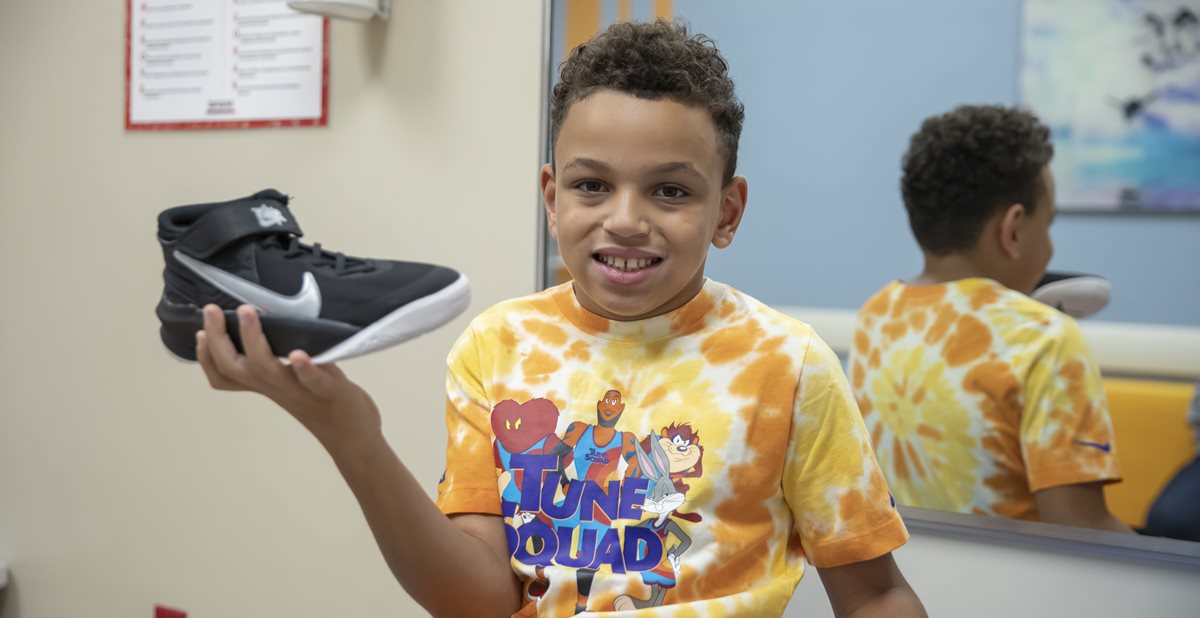
Most braces are built to fit in athletic shoes. They may also fit in hiking boots or lace-up dress shoes. Sometimes it can still be difficult to find properly fitting shoes. When buying shoes, always fit the shoes to the child with the brace on. Each brand and style of shoe fits differently. With a brace, most children will need wide shoes. Also, shoes with tongues that extend closer to the toe will fit braces more easily.

Shoes from Orthotics
When fitting a new leg brace, Scottish Rite for Children may provide one pair of athletic shoes. This allows us to see how the brace works and will allow your child to start wearing the brace immediately. These will not be replaced when they wear out.

Shoe Lifts or Wedges
Most children like to have several different pairs of shoes to wear, therefore you may want to have multiple pairs adjusted. A shoe lift or wedge can be added to shoes that you provide. For these buildups, drop off or mail the shoes to the Orthotics department. It may take several days to complete the work. Some shoes with silicone or gum rubber soles cannot be modified.

Modifying Shoes to Fit Braces
Certain shoes can be modified to allow them to fit better with a brace. It is important to make sure that the shoe comes close to fitting, before deciding to modify it.

  • Remove the insole.
  • Cut the tongue longer.
  • If the shoe is made of leather, a shoe repair shop can stretch the shoe.

Differently Sized Feet
Most children who wear a single brace, or have different sized feet, can wear the same sized shoes on both feet. Buy shoes to fit the larger. If the shoe is too loose on the smaller foot, try modifying that shoe.

  • Add an additional insole.
  • Pack the toe section of the shoe with cotton balls.
  • Put a dancer’s toe pad in the toe of the shoe.
  • Have your child wear two socks on the small side.

These modifications may not always work. If your child’s feet differ by at least two full sizes, you may need mismatched shoes. The easiest way to do this is to buy two pairs of shoes and discard the unneeded shoes. See the other side for additional resources.

Shoe Stores
A full-service shoe store can help fit your child with shoes.

Mismatched Shoes
If your child’s feet differ by at least two sizes, he or she may need mismatched shoes. Companies that sell single or mismatched shoes:

Services to share shoes between individuals with the opposite shoe concerns:

  • National Odd Shoe Exchange
  • Shoewap
  • Odd Shoe Finder

Special Shoes
Most children are able to wear standard shoes with their braces. However, you may need to special order an extra-wide style. Some models include:

  • BILLY Footwear (toe zippers)
  • Saucony Kids
  • Stride Rite
  • Piedro
  • Plae
  • Apis Mt. Emey
  • Nike FLYEASE (heel zippers)

Download the PDF.

Exercises for Hip Pain

Exercises for Hip Pain

Hip pain can be caused by a variety of underlying factors. Weak hip muscles can cause poor body mechanics when walking or performing other activities, which can eventually lead to pain. Muscles that are too tight may also cause pain and/or popping sounds in the hip during movement. Stretching and exercises can help improve the strength and flexibility of the hip muscles, decreasing pain and improving overall hip health.
 
Working with your child on proper stretching and exercises can help prevent hip pain. We recommend the following exercises and stretches to strengthen hip muscles:

Quadruple Fire Hydrant

  • Get down on all fours.
  • Keeping your knee bent, lift your hip out to the side.

Single-Leg Bridge

  • Lie on your back with knees bent.
  • Lift your hips off of the ground and then lift and straighten one leg.
  • Lower leg and repeat on the other side.

      Side-Lying Hip Abduction

      • Lie on your side with the bottom leg straight or bent.
      • Tighten the muscles on the top of your leg, in the front part of your thigh.
      • Lift the top leg while keeping the muscle tight and your knee straight.

          Half-Kneeling Hip Flexor Stretch

          • Kneel on one leg.
          • Slowly push your pelvis down while slightly arching the back until a stretch is felt on the front of your hip.

                      Supine Hamstring Stretch

                      • Lie on your back with one leg on the ground and one leg extended straight.
                      • Hook a strap on the extended leg to reach a maximum stretch.
                      • Straighten the extended knee further by tightening your front thigh muscles (quadriceps.)
                      • Slowly press the other leg down as close to the floor as possible.

                          Standing ITB Stretch

                          • Stand with one leg crossed behind the other leg.
                          • Bending at the waist, reach toward your back foot.

                              While daily exercises and stretches may prevent or alleviate hip pain, in some cases physical therapy might be needed. At Scottish Rite for Children, our Physical Therapy experts can work with your child to create a custom home-exercise program to address their specific needs. The home exercise program can be adjusted based on your child’s pain, progress and goals.

                              Learn more about our Physical Therapy services.

                              Stress Fractures in the Spine: Spondylolysis

                              Stress Fractures in the Spine: Spondylolysis

                              Pediatric orthopedic surgeon Jaysson T. Brooks, M.D., presented this as part of Coffee, Kids and Orthopedics education series. Brooks provided a detailed discussion of evaluating stress fractures in the spines of adolescents.

                              You can  and print the pdf.

                              watch the full lecture -What is Spondylolysis?

                              The facet joints in the back of the spine are connected by small segments of bone called pars interarticularis. Since this portion of the spine doesn’t get a great blood supply, it is at risk for stress fractures. This condition is called spondylolysis. Spondylolysis occurs more commonly at the L5 level and less commonly at the L4 level.

                              Most kids aren’t born with spondylolysis; it is caused by overuse and repetitive mechanical stress or forces. Activities or sports with repetitive hyperextension can cause a stress fracture of the spine. We see a higher incidence of spondylolysis in adolescents – as many as 47% of those with back pain. This is typically higher during growth spurts. The condition is much less frequent in adults. Some estimate 5% of adults with low back pain have spondylolysis.

                              In some cases, the stress fracture occurs bilaterally and the vertebra can slip forward, which is called spondylolisthesis. If a slipped vertebra presses on a nerve, it might cause severe shooting pain down the leg, and surgery may be required. However, if it breaks and doesn’t slip forward, surgery might not be necessary.

                              Spondylolysis: Genetic Predisposition?

                              • Spondylolysis occurs in 15-70% of first-degree relatives
                              • Prevalence
                                • White: 6%
                                • Black: 2-3%
                                • Indigenous American (Inuit): as high as 40%

                              History Matters

                              There is a higher prevalence of spondylolysis in elite athletes who report playing sports with repetitive hyperextension/rotation of the lumbar spine. Back pain should raise suspicion in these athletes:

                              • Football lineman
                              • Cheerleaders
                              • Gymnasts
                              • Weightlifters
                              • Divers / Swimmers

                              Back pain without a history of injury or repetitive activities is less likely to be caused by a stress fracture. In cases with shooting or decentralized pain, disc herniation should be considered.

                              Exam

                              The physical exam to assess for a stress fracture begins with palpation, and pain should be centralized around L5-S1 area. Active extension and hyperextension will be more painful than flexion. Coordination and strength should not be affected unless there is some nerve involvement, but pain may impact their ability to perform activities like heel walking and single leg hopping.

                              Imaging

                              In most cases, especially if the patient heard a “pop” and has acute low back pain, a standing anterior-posterior (AP) and lateral X-ray of the lower lumbar spine is recommended.

                              A study published in the Journal of Pediatric Orthopaedics looked at 2,846 patients with a median age of 14.6 years that were seen for back pain. 76% had no clear cause for their back pain, and less than 61% had two or fewer follow-up visits. This is a good reminder that not every patient with back pain has a stress fracture.
                              X-rays may not show early signs of spondylolysis. Rather than automatically ordering advanced imaging, a pediatric sports or spine referral may be the best next step because MRIs may also be inconclusive.

                              Treatment

                              Treat conservatively first.

                              • Activity Modification: 3 – 6 months
                              • Physical Therapy: 3 – 6 months
                                • Focus on core strengthening to improve lumbar stability
                              • Non-steroidal anti-inflammatory drugs (NSAIDS)
                                • Meloxicam and/or diclofenac cream
                                • Naproxen
                              • Bracing may provide comfort but does not affect return to activities.

                              Often patients only want to do one of these, but that may make extend their recovery by several months.
                              It is acceptable if a fracture never heals on an X-ray as long as the symptoms go away. If six months of conservative treatments only show slight improvements, a pars injection may help their symptoms. Some patients are injected every six months.

                              Surgery should always be a last resort.
                              If the gap is not too wide, a screw is used for a direct pars interarticularis repair. A fusion of the surrounding vertebra may be considered if a loss of motion is acceptable.

                              Check out our latest on-demand lectures available for medical professionals.

                              Infants and Developmental Dysplasia of the Hip

                              Infants and Developmental Dysplasia of the Hip

                              This article was originally published in the Pediatric Society of Greater Dallas newsletter. Committed to improving orthopedics care of pediatric patients in all settings, Scottish Rite for Children specialists are regular contributors to this publication for local pediatricians in North Texas.

                              Developmental dysplasia of the hip (DDH) is the most common orthopedic condition affecting newborns. The overall incidence has been estimated at approximately 1%. Dysplasia is a term that means poorly formed. It describes this condition well because one or both sides of the hip joint do not grow correctly as the child develops. In severe forms of DDH, the hip joint can be completely dislocated, meaning that there is no contact between the ball of the hip joint (femur) and the socket (acetabulum). 

                              Print the PDF.

                              Screening for DDH
                              The American Academy of Pediatrics (AAP) published a clinical report on current standards for evaluating and treating DDH. With later recognition of the condition, the treatment becomes more complex and may even require surgery. In order to minimize missed cases of hip dysplasia, the AAP recommends that pediatricians periodically screen for DDH during routine office visits, from infancy until the child is walking. With effective screening, most cases are identified and managed during infancy, leading to complete correction of hip dysplasia and the development of normal hips.

                              Ann,-Hip.jpg

                              As a pediatric orthopedic surgeon, Corey S. Gill, M.D., M.A., cares for many children with DDH and has received several questions from referring providers about appropriate care. The most important things for pediatricians and other referring providers to understand about DDH include:

                              • Perform a hip examination on every newborn and infant patient. Soft tissue clicks around the hip and knee are very common and do not generally indicate hip dysplasia. Similarly, asymmetric skin creases on the inner thigh do not usually mean there is a problem with the hip. Findings that are clearly abnormal and should lead to orthopedic referral include:
                                • An unstable hip that “clunks” into or out of place. Hip stability is evaluated during the exam by performing the Barlow and Ortolani maneuvers. The Barlow test identifies a hip that is in place but can be easily dislocated with gentle pressure. The Ortolani test identifies a hip that is dislocated at rest, but can be placed back into the joint with positioning of the thigh.
                                • Significantly decreased or asymmetric range of motion. This is especially important for abduction of the hips, which is moving the hips out to the side when lying down. Differences as small as 10 degrees compared to the normal side may indicate a significant problem.
                                • A significant leg length difference, which may indicate a hip dislocation. Leg length difference is best evaluated with a Galeazzi test. This test is performed by flexing the hips to 90 degrees and checking to see if the knees are level.
                                • In toddlers and older children, decreased hip abduction and a waddling gait, limp or unilateral toe walking may indicate hip dysplasia or dislocation.
                              • Identify the risk factors that make hip dysplasia more likely. The two most important are family history of hip dysplasia and breech presentation (especially frank breech). Providers should have a low threshold for orthopedic referral in these patients. Other risk factors include female sex, first born child and oligohydramnios.
                              • Understand the right time to refer a patient for DDH evaluation. In newborns with unstable hips on exam, a referral should be made immediately so treatment can start as soon as possible. In children with a normal exam but risk factors for DDH, an ultrasound should be obtained at approximately six weeks of age. Obtaining an ultrasound in children earlier than this often leads to a false positive diagnosis of DDH secondary to physiologic immaturity of the hip joint in the newborn.

                              Orthopedic Intervention

                              When infants do need orthopedic intervention for hip dysplasia, our first line of treatment is a Pavlik harness. This fabric and Velcro harness is generally worn for 23 hours per day for approximately six to eight weeks, but it is removable for bathing. The harness keeps the hips flexed and rotated in the correct position for normal development of the hip joint. After treatment with a Pavlik harness, we use physical exams, ultrasound and X-rays to monitor growth and confirm the hip joint is developing normally. Most infants with DDH require no further orthopedic treatment after wearing a Pavlik harness.

                              In some infants, especially those with severe hip dysplasia or a dislocation, Pavlik harness treatment may not be successful. Occasionally, a different type of hard plastic brace may be successful in correcting the hip dysplasia in these children. However, most children who do not respond to Pavlik harness treatment will ultimately require surgical intervention to prevent long term problems from hip dysplasia such as cartilage injury, limp, leg length difference and early arthritis. Depending on the severity of the hip dysplasia, surgical treatments may include:

                              • Closed reduction – This involves repositioning the ball of the hip joint deeply into the socket when the child is asleep under anesthesia and then applying a body cast called a spica cast for a total of three to four months. During this procedure, we often inject a small amount of medical dye into the hip joint to confirm that the ball of the hip joint is appropriately positioned in the socket. This is called an arthrogram.
                              • Open reduction – Sometimes the hip joint will not line up well with repositioning of the leg because there are tight tissues blocking the ball from sitting deeply in the socket. In these cases, an incision is made in front of the hip where the tight tendons, ligaments and soft tissues are moved out of the way. Afterwards, the lining of the hip joint is tightened with a strong suture to help hold the hip in position. This procedure is called a capsulorrhaphy.
                              • Osteotomies – In older children (over age 1.5 – 2 years), soft tissue procedures alone are often not enough to ensure the hip joint is lined up well. In these cases, we often supplement the open reduction procedure by cutting the bone in a controlled way to help reorient the hip into the socket. This is called an osteotomy and can be performed on the ball side of the hip (femur osteotomy) or socket side of the hip (pelvic osteotomy). Metal implants are often used to hold the bone in the new position and are removed at a later date.

                              Conclusion

                              Hip dysplasia is a common orthopedic condition in newborns that can lead to significant long-term consequences if left untreated. Certain risk factors such as family history of dysplasia and frank breech presentation greatly increase the risk of developing DDH. Pediatricians play a crucial role in examining infants, identifying those with risk factors and referring them to a pediatric orthopedic specialist when appropriate. When diagnosed in the first few months of life, noninvasive treatment with a harness or brace is highly successful and generally leads to the development of a normal hip. In some cases of severe hip dysplasia/dislocation or in cases of delayed diagnosis, surgical intervention is required to improve the long term prognosis of the hip joint.

                              Referral Tips 

                              A potential diagnosis of hip dysplasia can lead to significant anxiety for new parents. Understanding the best time to refer patients and initiate treatment helps to maximize treatment success and efficiency while minimizing parental stress and worry.

                              • For infants with risk factors for DDH such as family history or breech presentation but a normal physical exam, an ultrasound should be obtained around six weeks of age. Ultrasounds performed earlier than this age result in a large number of false positives and potential unnecessary treatment in a harness.
                              • There is no need to obtain an ultrasound prior to referral as we work closely with experienced ultrasound technologists who can perform the diagnostic hip ultrasound on the same day as an infant’s office visit.
                              • In children with a clearly abnormal exam (unstable/dislocatable hip or asymmetric hip abduction) in the nursery or in routine office visits, immediate referral should be made so that treatment in a harness can be initiated as soon as possible. In these children, there is no need to wait until the child is 6 weeks of age for referral.
                              • If only abnormal exam finding is a “hip click” or asymmetric thigh crease, referral and ultrasound should be deferred until 6 weeks of age given the relatively low prevalence of DDH in these children.
                              • In premature infants still in the NICU with risk factors for DDH, it is generally OK to wait for referral until after the child is discharged to go home. If an examiner finds the hip to be unstable while still an inpatient, phone consultation with a pediatric orthopedic surgeon is available to answer questions or discuss the most appropriate time to see the patient.
                              • If a family has an infant diagnosed with DDH, all future siblings of the child should be referred for screening, ultrasound at six weeks of age and strong consideration should be given for referral of older siblings for a hip radiograph. First degree relatives have more than a tenfold higher risk of DDH compared to controls.