Muscle Strain Q&A

Muscle Strain Q&A

Our world-renowned sports medicine experts are ready to help your injured athlete get back in the game. We have unparalleled experience providing nonoperative and arthroscopic care to treat common sport-related injuries including concussions, ligament injuries and cartilage conditions in the knee, ankle, shoulder, elbow and hip in young and growing athletes.

Sports Medicine expert Jacob C. Jones, M.D., RMSK, shares information about muscle strains and how to handle these types of injuries in young athletes.

What is a muscle strain?
A muscle strain is a disruption of the muscle fibers in a certain muscle group. Muscle strains can be mild or they can be severe, causing muscle tearing.

What causes a muscle strain?
Muscles are constantly being pushed and pulled, but when a muscle contracts at the same time that it is being pulled, a strain can occur. This type of muscle movement is called an eccentric contraction. 

What are the symptoms of a muscle strain?
In mild strains or low-grade muscle disruptions, the most common symptom will be pain in the area. Severe disruptions or tears can also cause swelling, more noticeable weakness, and even bruising.

Should you seek medical treatment for a muscle strain?
It is definitely wise to seek medical treatment for muscle strains. In mild cases, a young athlete may want to consult with their athletic trainer for advice and recommendations on reducing the pain. Athletic trainers can also help determine whether the athlete needs to see a physician for the injury.

Relative rest, in combination with muscle rehab, is the best treatments for a strain. It is important to allow the muscle to heal while also building strength and flexibility to avoid further injury. Even in high grade muscle tears, surgery may not be commonly recommended. 

Are certain muscles more at risk for strains?
Yes, muscle groups that are at the highest risk for strains are those that cross multiple joints. For example, some hamstring and quadricep muscles cross both the hip and knee joints and calf muscles cross the ankle and knee joints. Any muscle can be strained, but those groups are more likely to be injured.

How can you avoid muscle strains?
Muscles are less likely to have a strain if they are flexible and strong. Stretching daily can help provide your muscles with more flexibility and strength. Additionally, it is important to also warm up your muscles before working out or playing a sport. Muscles are less likely to strain or tear when they are warm, so it is important to not skip warm-ups before practice.

What does recovery from a muscle strain look like?
Once pain allows, it is important to do some rehabilitation to the muscle before returning to regular activity. In mild strains or low-grade disruptions, recovery time may take weeks. In more severe cases that lead to muscle tears, recovery time may take months. We look for good range of motion, minimal to no pain, and good strength prior to return to sport.

What happens if an athlete returns to sports or activity before the strain is healed?
The biggest risk of returning to athletics or sports too soon is re-aggravating the muscle and extending the recovery time. Additionally, having a strain may cause you to favor one leg or arm and could lead to further injury.

How can ultrasound be used to diagnose and treat muscle strains?
Specially trained experts can use musculoskeletal ultrasound to evaluate injured joints, ligaments, tendons, muscles and bones. Ultrasound can visualize soft tissues like muscle well with a high level of detail. When looking at a muscle using ultrasound, a low-grade strain may show some edema, swelling caused by fluid in tissue, while a more severe strain that has already torn will clearly be visible. Using ultrasound can also allow physicians to determine where additional treatment or care is needed in treating muscle strains. Ultrasound can also be used for treatment of chronic muscle tears not improving with other conservative measures.

Sports medicine is a medical and surgical specialty that considers the comprehensive needs of athletes and provides management for sport-related injuries and conditions. Young and growing athletes are highly competitive and have unique conditions that require care by a pediatric team of experts. Learn more about our Center for Excellence in Sports Medicine and how board-certified pediatricians, pediatric orthopedic surgeons, physical therapists, athletic trainers, psychologists and other sports medicine specialists work side-by-side with each athlete, their parents and coaches to develop the best game plan for treatment, rehabilitation and safe return to sport.
D Magazine: How Collin County’s Healthcare Providers Are Managing Record Population Growth

D Magazine: How Collin County’s Healthcare Providers Are Managing Record Population Growth

Collin County is the fourth-fastest growing county in the country. Growth can present all sorts of challenges for professionals working in education, healthcare, transportation and every other area of public life.

Philip L. Wilson, M.D., was part of a panel of Collin County leaders, talking about how their organizations met the challenge of keeping people healthy in the midst of a population explosion. 

Read the full article. 

Study Looks at Re-Injury Rate After ACL Reconstruction in Young Athletes

Study Looks at Re-Injury Rate After ACL Reconstruction in Young Athletes

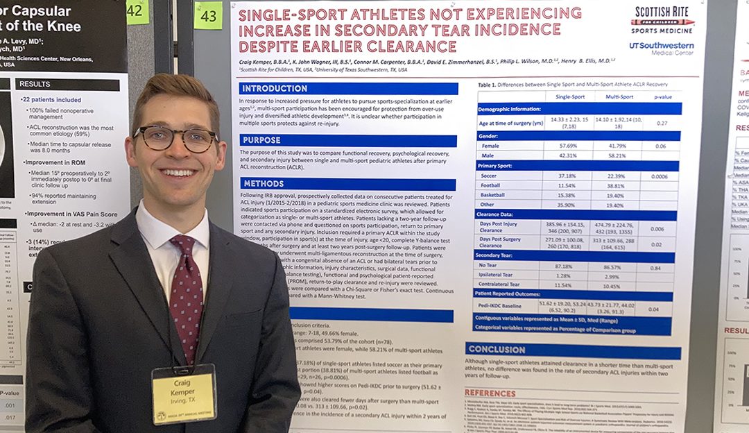
Wrapping up his third year as a medical student at UT Southwestern Medical School, Craig Kemper, B.B.A., has participated in several projects with the Center for Excellence in Sports Medicine research team. Kemper was the lead author on a project looking at athletes who were back to sport after an anterior cruciate ligament (ACL) tear and reconstruction that was recently presented at the 39 annual meeting of the Mid-America Orthopaedic Association. The organization comprises orthopedic surgeons from 20 states, including Texas.

After surgery for the “primary” ACL tear, as many as 1 in 4 young athletes re-tear the reconstructed ACL or the ACL in the other knee. The rate for these “secondary” ACL tears in young athletes is a concern for researchers and clinicians in pediatric sports medicine. This review included patients seen over three years at Scottish Rite for Children for an ACL tear and reconstruction to determine whether participation in multiple sports protects against re-injury.

The 145 patients in the study were

  • an average age of 14 years.
  • 50% male, 50% female.
  • > 50% reported playing only one sport (single-sport athletes)
    • Most played soccer.
    • On average, these athletes returned to sports in fewer days than multi-sport athletes.

Kemper says young athletes continue to feel pressured to choose one sport at earlier ages to “not be left behind.” Many recommend multi-sport participation to help an athlete develop varied skills and protect from overuse injuries unique to growing children, including apophysitis and osteochondritis dissecans.

“We give this advice, but we aren’t sure if it applies to this population regarding re-injury after an ACL reconstruction,” says co-author and Medical Director of Clinical Research, Henry B. Ellis, M.D. “The time out of sports is already so long for an ACL tear, we are eager to learn all the variables that contribute to re-injury rates. Other studies have looked at surgical techniques, but this one looks at sport-participation and time to return-to-play.”

Although single-sport athletes were cleared to return to sports in a shorter time than multi-sport athletes, the analysis found no difference in the rate of secondary ACL injuries within two years of follow-up for this group. “Results like this are still helpful and give direction for future projects,” says Ellis. “More importantly, they help me as a pediatric orthopedic surgeon know how to counsel my patients.”


SINGLE-SPORT ATHLETES NOT EXPERIENCING INCREASE IN SECONDARY TEAR INCIDENCE DESPITE EARLIER CLEARANCE, Craig Kemper, B.B.A., K. John Wagner, III, B.S., Connor M. Carpenter, B.B.A., David E. Zimmerhanzel, B.S., Philip L. Wilson, M.D., Henry B. Ellis, M.D.

Learn more about ACL injuries on our website.

Share Your Story: A Happy Ending

Share Your Story: A Happy Ending

Meet Max, a patient seen by our experts in the Center for Excellence in Hip. Learn more about his journey below.

Blog written by Max’s mom, Melinda. 

Giving birth is one of the most exciting things a mother can experience. When our son, Max, was born, he weighed 10 pounds. We couldn’t wait to bring him home from the hospital, so we could begin getting to know him. When the pediatrician visited him in the hospital for his newborn exam, he identified that Max had symptoms of hip dysplasia. Developmental dysplasia of the hip occurs when a baby’s hip socket is too shallow to cover the head of the thighbone properly, and left uncorrected, he might experience pain when he tried to learn to walk.

Fortunately, our pediatrician recognized the issue and recommended we take Max to Scottish Rite for Children for treatment. The pediatrician’s office made our initial appointment, and Max had his first consultation when he was just a week old. As first-time parents, there was a lot we didn’t know about taking a baby to a doctor’s appointment. For example, we didn’t even pack a diaper bag because we thought we’d be home long before he needed a diaper change. 

My husband and I were very nervous — we knew nothing about hip dysplasia, and we were worried about the long-term consequences. Could it be fixed? Would he eventually be able to walk? The team at Scottish Rite and our pediatric orthopedic surgeon Dr. John Birch were fantastic. They calmed our fears and answered all of our questions. During the comprehensive evaluation, they provided a private room for us to use when Max needed to nurse. They also provided us with diapers when he needed to be changed. He was a big baby — apparently his newborn diapers and T-shirt were already too small, so they gave him a new T-shirt too. 

Max was fitted with a Pavlik harness that he wore 24 hours a day, except during bath time. We visited Scottish Rite weekly for several months. The doctors assessed his progress and adjusted his harness as he grew. The doctors and staff could not have been nicer or more supportive. After a couple of months, they determined that his hips had grown and developed properly, and he would no longer need to wear his harness. We returned to Scottish Rite regularly to gauge his progress. He started walking at 14 months, without any problems. We eventually moved to San Antonio but returned to Scottish Rite annually to make sure his hip and leg development was on track. We couldn’t have been happier with the support and guidance we received. 

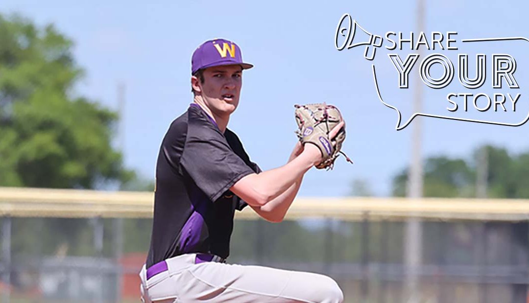
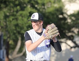
Everyone loves a happy ending, so we’re happy to share ours. Max grew and grew – he played varsity basketball and baseball during high school. He recently enrolled at Williams College in Massachusetts, where he is continuing his baseball career. He’s a 6’8 left-handed pitcher, and he can’t wait to play for the Ephs. We are incredibly grateful to the team at Scottish Rite for the compassion and amazing care we received. 

DO YOU HAVE A STORY? WE WANT TO HEAR IT! SHARE YOUR STORY WITH US.

Cerebral Palsy: Defining the Common Terms

Cerebral Palsy: Defining the Common Terms

Cerebral palsy (CP) is a condition that affects posture, movement and balance associated with an injury to the developing brain. The injury is static (does not worsen) and occurs before or during infancy. Scottish Rite for Children provides a multidisciplinary approach to care for children diagnosed with CP. Below are the most common terms used when our team is talking with patients and families about the condition. 

Motor Difference

  • Hypertonia – Increased muscle tone or a tendency for muscle to be tight.
  • Hypotonia – Low muscle tone, or a tendency for muscles to be excessively relaxed.
  • Dyskinesia – Excessive muscle movements.
  • Spasticity – Muscle tightness that manifests as a catch and release when a limb is moved quickly about a joint. This type of muscle tightness is constant and consistent throughout the day.
  • Dystonia – Muscle tightness that occurs because of unintended muscle activation. This type of muscle tightness can occur when an individual is trying to move other body parts or with certain emotions.
  • Ataxia – Motor pattern that describes difficulties with balance and difficulty with performing smooth limb movements.

Anatomic Involvement

  • Diplegic cerebral palsy – This term describes a motor difference that involves both legs.
  • Hemiplegic cerebral palsy – This term describes a motor difference that involves one side of the body and including the leg and arm on the same side.
  • Quadriplegic cerebral palsy – This term describes a motor difference that involves all four limbs.
  • Triplegic cerebral palsy – This term describes a motor difference involves both legs, and one arm.

Motor Classification

  • Gross Motor Functional Classification System (GMFCS) – This is a system used by clinicians to categorize how a person with CP moves and functions in different environments. It helps clinicians come up with treatment plans and anticipate changes that can occur in the body as the individual ages.
  • GMFCS I – Individuals in this category of mobility are able to go walk around their environment with little to no help. They can go up and down stairs without holding on to a handrail.
  • GMFCS II – Individuals in this category of mobility may need more help when walking on uneven surfaces or inclines and can go up and down stairs by using a handrail.
  • GMFCS III – Individuals in this category of mobility use an assistive device to walk community distances, such as a walker or crutches. They may use a wheelchair for longer community distances
  • GMFCS IV – Individuals in this category of mobility use a wheelchair for most of their mobility. They may be able to propel manual wheelchairs without any help, and they require little to no head and trunk support when sitting.
  • GMFCS V – Individuals in this category of mobility use the wheelchair for all of their mobility. They may be able to drive a motorized wheelchair and typically need head and trunk support when sitting.

Related Neurologic Findings

  • Periventricular Leukomalacia – This is a pattern that is seen on brain imaging that indicates scarring around the ventricles (the area of the brain that hold spinal fluid). It is usually associated with injury to the vessels around the ventricles in premature infants and affects both sides of the brain.
  • Hydrocephalus – This is a finding on imaging that indicates that the ventricles are enlarged.
  • Porencephaly – This is a pattern on brain imaging that indicates a local injury to a specific area of the brain. It usually affects only one side of the brain.

Related Orthopedic Diagnoses

  • Contracture – This describes a limitation in range of motion at a joint. There are multiple factors that contribute to contractures.
  • Hip migration – Tendency for the femur bone to become uncovered by the hip bone. This can occur in individuals with weakness or increased tone.
  • Neuromuscular scoliosis – A curvature of the spine that is related to weak or spastic muscles.