Adolescent Hip Dysplasia and Other Causes of Hip Pain

Adolescent Hip Dysplasia and Other Causes of Hip Pain

Content included below was presented at the 2021 Pediatric Orthopedic Education Symposium by pediatric orthopedic surgeon William Z. Morris, M.D.

You can watch the full lecture and download this summary.

In hip dysplasia, the acetabulum (or hip socket) is shallow and doesn’t adequately cover the femoral head. Developmental dysplasia of the hip (DDH) occurs in approximately 1% of newborn children, and it is associated with four risk factors:

  • Female
  • Firstborn
  • Feet first (breech)
  • Family history

Hip dysplasia is relatively more commonly diagnosed in skeletally mature adolescents, affecting around 3% to 5% of the asymptomatic population. Cross-sectional studies have shown that female sex and a family history of dysplasia remain risk factors in adolescents.

There has been growing attention to the treatment of hip dysplasia as there is an association between hip dysplasia and the development of early osteoarthritis. In 1939, Gunnar Wiberg first described hip dysplasia and objectively measured it using what is now called the lateral center edge angle, to describe how well the socket (acetabulum) covers the ball (femoral head). On an AP (anterior posterior) pelvis X-ray, the angle is created by a vertical line through the center of the femoral head and a line from the center of the femoral head to the lateral border of the acetabular fossa. A larger angle reflects greater hip coverage, and a smaller angle reflects less coverage, commonly seen in a dysplastic hip.

Wiberg followed patients with dysplasia for up to 30 years and found that all of the patients with hip dysplasia eventually developed osteoarthritis. The smaller their center edge angle was (reflecting greater hip dysplasia), the faster they developed osteoarthritis.

Development of Osteoarthritis in Dysplastic Hips
The development of early osteoarthritis is suspected to occur due to a couple mechanical reasons. The most important factor is that:

Pressure = Force / Unit Area
In the hip, pressure on the joint surfaces depends on the total surface area of the femoral head in contact with the socket. A well-covered femoral head distributes weight-bearing forces across a larger surface area, reducing the pressure on each unit of cartilage. In contrast, a dysplastic acetabulum offers less surface area which increases the pressure on the cartilage contributing to earlier hip degeneration. In addition to the smaller weightbearing surface, the acetabulum is also more obliquely oriented. Therefore, compressive forces are less and shearing forces are greatly increased. This increased shear force may also contribute to cartilage degeneration.

Symptoms of Adolescent Hip Dysplasia
There are many different causes of hip pain in an adolescent patient and combining clues from the history and physical exam is essential to determine the underlying problem. The location of a patient’s pain can help determine the underlying etiology. Intra-articular pain of the hip usually presents as anterior groin pain, often due to a cartilage injury, a tear in the labrum (the ring of cartilage at the periphery of the socket) or inflammation of one of the hip flexors called the Iliopsoas tendon. Lateral hip pain that locates over the greater trochanter (the bony prominence on the lateral aspect of the thigh) probably reflects trochanteric bursitis or inflammation of the bursa overlying that region. Pain or soreness after activity above the greater trochanter where hip abductors like the gluteus medius are located may signal hip abductor fatigue, which is common with hip dysplasia. Pain over the iliac crest where the abdominal musculature attaches could reflect some inflammation of that apophysis, of the anterior superior iliac spine (ASIS) where the sartorius attaches, or of the anterior inferior iliac spine (AIIS) where the rectus tendon attaches.

Additional Factors to Consider when Discussing Hip Joint Symptoms

  • Pain
    • Duration
    • Aggravating factors
      • Squatting, stairs, low chairs
    • Alleviating factors
  • Mechanical symptoms
    • Locking, popping, catching, snapping
  • Neurological symptoms (which may suggest spinal pathology)
    • Radiating pain, paresthesias

Physical Exam

  • Pain Assessment
    • Palpation can reproduce symptoms and help to localize the pain.
  • Strength Testing
  • Range of Motion
    • Limited internal rotation (IR) can indicate hip impingement or a more acute concern, slipped capital femoral epiphysis (SCFE)

Special Tests

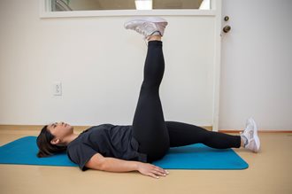
Straight Leg Raise Test
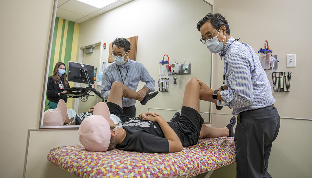
A straight leg raise is a passive test that helps to distinguish between hip and spine pathology and is performed by flexing the hip with an extended knee in a supine position. If this maneuver reproduces the patient’s pain that radiates distally, the problem may be related to nerve compression in the spine rather than a problem in the hip.

Trendelenburg Test

The hip abductors (e.g. gluteus medius) are typically weaker when patients come in with pain, so it can be targeted in physical therapy. The Trendelenburg sign is a quick physical examination used to assess for hip abductor weakness.
The patient stands on one leg (stance leg) and bends the other knee about 90°. Observe for evidence of hip abductor (i.e. gluteus medius) weakness which includes:

  • Pelvis drop contralateral to the stance leg.
  • Trunk lean/shift toward the stance leg.

Apprehension Test
Another test to further evaluate for dysplasia is the apprehension test:

  1. The patient lays in a lateral position
  2. Abduct the patient’s leg 30° away from midline
  3. Flex the patient’s knee 90°
  4. Gradually extend the hip

Patients who have an anterior uncovering of the socket from dysplasia will feel pain or a sensation of apprehension, which suggests that there may be some instability or dysplasia.

Femoroacetabular Impingement Test
Hip impingement should also be tested:

  1. Flex the hip to 90°
  2. Abduct the hip, bringing it towards midline
  3. Internally rotate the hip

This test attempts to reproduce hip impingement where the femoral head or neck collides against the socket. If this causes pain, there may be a cartilage injury such as a labral tear. An MRI is recommended to evaluate intra-articular soft tissues.

Slipped Capital Femoral Epiphysis – A condition not to be missed
Slipped capital femoral epiphysis (SCFE) is an adolescent disorder in which the growth plate is damaged and the femoral head epiphysis moves, or slips, with respect to the rest of the femur. Diagnosis in a timely manner is essential to prevent further injury to the hip.

Consider SCFE if these signs are present:

  • limp
  • walk with their foot externally rotated
  • have limited range of motion, especially with internal rotation

Obligate external rotation is nearly pathognomonic for slipped capital femoral epiphysis, so if the patient can’t flex their hip straight up without turning their leg into an externally rotated position to accommodate further flexion, an anterior-posterior and frog pelvis film is recommended to ensure slipped capital femoral epiphysis hasn’t been missed. Immediate non-weight bearing with a wheelchair and urgent referral to pediatric orthopedics or an emergency room is recommended for this condition.

Radiographic Evaluation
A radiographic evaluation is important for a definitive dysplasia diagnosis. The AP pelvis film is a workhorse tool for the evaluation of hip coverage. It is taken standing to allow assessment of the patient’s hip coverage in their functional position using the lateral center edge angle (LCEA). A hip with an LCEA less than 25° is considered dysplastic.
Imaging also allows an assessment of the inclination of the socket. By drawing a line between the medial and lateral edges of the roof of the socket and measuring the angle between that line and a horizontal line, physicians can determine the acetabular inclination. The more inclined the socket is, the more dysplastic.

Treatments
Treatment for dysplasia begins with nonoperative options, which include:

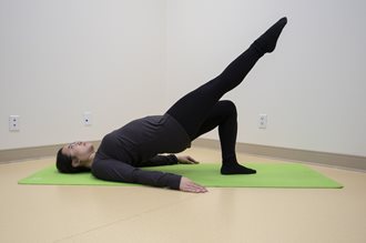
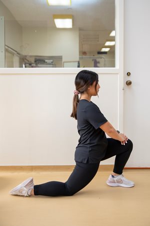
  • Physical therapy
  • Activity modifications
  • Non-steroidal anti-inflammatory drugs (NSAID)

When nonoperative treatments don’t work, and patients continue to have radiographic dysplasia and pain, including abductor fatigue pain above the greater trochanter or anterior intra-articular groin pain, they are treated surgically with a periacetabular osteotomy (PAO). This specialized procedure is done in patients who are approaching skeletal maturity or are already skeletally mature. Several cuts are made around the socket of the acetabulum to mobilize the socket. The socket is then reoriented to better cover the femoral head.

Outcomes
For patients with symptomatic hip dysplasia, the PAO has been shown to be successful in improving patients’ function, getting them back to their activities/sports, and preventing early hip replacement. In patients with hip pain and clinical or radiographic evidence of acetabular dysplasia, please consider a referral to Scottish Rite for Children for discussion of the condition and shared decision-making in the plan for management.

Check out our latest resources for medical professionals
Checking In – Perthes Disease

Checking In – Perthes Disease

Checking In – A Scottish Rite for Children Podcast, Episode 4
Perthes Disease

Host: Clinical Nurse Manager, Jennifer Bowden, R.N.
Expert Guests: Director of the Center for Excellence in Hip and pediatric orthopedic surgeon Harry Kim, M.D., M.S., and special guest Kristen Odom, R.N.

Listen to the full episode.
Below are the highlights from this episode:

What is Perthes disease?

Perthes disease is a pediatric hip disorder that usually affects children between the ages of 2 and 14.  The cause of this condition is currently unknown. Perthes disease leads to a loss of blood flow to the hip joint; however, experts do not know why that blood flow is disrupted.

What are some important terms that parents should always be aware of or understand when talking about this condition?
Perthes disease affects the hip. The hip joint includes a ball (femoral head) and socket (acetabulum). Perthes affects the ball part of the hip, causing a disruption and loss of blood supply, which initiates the disease. This leads to necrosis, or death of the bone. A common term used when talking about Perthes is osteonecrosis – osteo=bone and necrosis=death, meaning bone death.

What are the different stages of Perthes disease? 

There are four stages to the disease.

  • The first stage is when the blood flow gets disrupted, also known as necrosis – the stage of bone death.
  • The second stage is fragmentation which is when the body tries to heal the bone, but the ball (femoral head) is weakened, causing it to break down. Because the body is also trying to remove the dead bone, the head starts to collapse or flatten out, which causes it to lose its round shape.
  • The third stage is re-ossification which is when the body begins to remove the dead bone and start to build back the new bone.
    • Re-ossification: 
      • Re: is the starting back
      • -ossi: is the bone
      • -fication: is the new remaking of the bone
  • The fourth stage of Perthes disease occurs when the hip joint is all healed, also known as the healed stage.

What are the symptoms of Perthes disease? 

  • Persistent limping.
  • Pain in the hip, knee or thigh.
  • Stiffness in the hip and/or decrease in range of motion.

How is Perthes diagnosed? 

The diagnosis of Perthes requires determining that the cause of pain is not due to other issues or another condition. A thorough history is taken to make sure it is not something else, and then an examination is done to assess the hip as well as the knee to make sure it is the hip that’s the problem. The diagnosis of Perthes requires X-rays because it’s not just a clinical diagnosis. Diagnostic imaging is required.

For older children, patients over the age of 6, our team usually orders a specific MRI called a perfusion MRI. This test can assess how severe the disease is and provides more clarification on if operative treatment would be beneficial/necessary for the child.

What makes the perfusion MRI an important part of diagnosing Perthes disease?

The perfusion MRI involves injecting MRI contrast into the hip so that the blood flow can be analyzed to better understand where blood flow is lacking. More loss of blood flow means that the disease is more severe, and the healing process can take longer.

Why is it important to be seen by a pediatric orthopedic specialist for Perthes?

Perthes disease is a very uncommon condition that few doctors have experience in diagnosing and treating. It is important for parents to find a pediatric orthopedic specialist with a special interest in Perthes disease that cares for children with the condition regularly.

What can a parent and patient expect when they come to Scottish Rite to be seen for Perthes? 

The team at Scottish Rite provides a comprehensive assessment of the child – thorough history and physical exam, required imaging and reviewing any previous X-rays or testing that has been done previously.

The child’s emotional state is also assessed. They are asked to complete a PROMIS questionnaire which asks the child about anxiety, depressive symptoms and their peer relationship, as well as psychological aspects of the patient. Perthes is a chronic condition meaning the treatment can take months and, for some, years to overcome. Our team provides care for the whole child – mind, body and spirit.

What are the treatment options for Perthes disease?

All children are different, as is the severity of each child’s condition, so the treatment plan created for each child diagnosed with Perthes is also unique. Our team evaluates each patient – the stage of the disease they are in, range of motion and pain – then develops an individualized treatment plan.

Non-Operative Treatment

  • Weighted relief treatment – the child is given crutches, a walker or wheelchair to decrease the amount of weight placed on the hip.
  • Petrie casting – a treatment started back in the 1970s which involves putting both legs in the cast with a bar or two bars in between to spread the legs. This allows the hip to rest, especially for the children who are very active and unable to rest themselves.

Operative Treatment
Depending on the child and severity of their condition, surgery is an option to treat Perthes.

International Perthes Study Group
Scottish Rite for Children is the leading center for the International Perthes Study Group (IPSG). IPSG includes over 50 pediatric orthopedic surgeons and researchers from 10 different countries who are dedicated to improving the care of patients with Perthes disease.

The research from this group analyzes the very basic scientific level of the condition to better understand the disease and the key processes that are contributing to the femoral head collapsing and not healing properly. Through that research, the team is trying to develop new and innovative treatments to improve healing for children diagnosed with Perthes.

Learn more about the International Perthes Study Group.

NBC DFW: 8-Year-Old Battling Rare Disease Returns to Dallas for Treatment, Is Special Guest at Cowboys Game

NBC DFW: 8-Year-Old Battling Rare Disease Returns to Dallas for Treatment, Is Special Guest at Cowboys Game

Gavin Miller of Charlotte, Michigan visited Texas Scottish Rite Hospital for Children to see Dr. Harry Kim, the Director for Center for Excellence in Hip, for the ongoing treatment of his Perthes disease.

Miracle Flights, which provides children and their families free flights to distant and specialized care, flew Gavin and his mother to Dallas. Gavin was also able to see his “hometown hero” Cooper Rush with the Dallas Cowboys play in the team’s first preseason home game. Rush is also a native of Charlotte, Michigan and has followed Miller’s medical journey.

Watch NBC DFW’s feature or learn more about Perthes disease

Recognizing Adolescent Hip Conditions

Recognizing Adolescent Hip Conditions

Key messages from a presentation by staff orthopedist, David A. Podeszwa, M.D., at Coffee, Kids and Sports Medicine. Article originally published in first quarter, 2018 issue of Pediatric Society of Greater Dallas newsletter. 

Watch the lecture
Print the PDF

Recognizing Hip Conditions in the Pre-Teen and Teenager

Kids of all ages complain frequently of aches and pains around the hip and it is really easy to brush them
off. I would be lying if I said that I haven’t done it to my own children. For the super-active child/teen who participates in high impact activities year-round, it is easy to explain away complaints of hip pain as simple overuse. The combination of anti-inflammatories, stretching and playing through the pain is a common remedy. At the opposite end of the spectrum is the video gamer or book lover who is more sedentary and less interested in exercise. Their complaints of hip pain are easily attributed to deconditioning and weakness. Becoming more active is the simple remedy. Unfortunately, not all hip pain can be ignored. Missing certain conditions early in their presentation can have significant long-term pain and functional consequences. Below are several important pearls to remember that will help you avoid missing a serious hip condition when evaluating a patient with hip pain.

  1. Hip disorders can present with hip or knee pain. Sorting out the etiology and location starts with a good history and physical exam. Is the chief complaint pain, limp, or decreased motion? Some disorders can present without pain and only a limp. Where does it hurt? Hip disorders can present with hip (anterior, lateral, groin), thigh or knee pain. Complaints of constant pain that does not resolve with rest, is worse with weight bearing, limits hip range of motion, and is not improved with anti-inflammatories should be red flags for a significant underlying condition. Physical exam may demonstrate pain with palpation at the anterior superior iliac spine, iliac crest, and or greater trochanter. Pain with range of motion or significant asymmetry in hip range of motion should also be concerning.
  2. Children and adolescents do not get “groin pulls.” Recurrent limping and/or hip pain (especially groin pain) unresolved with rest is likely to have an underlying etiology. “Groin pull” is an easy answer, but it is never the correct one.
  3. An adolescent limping with his/her foot turned out and complaining of hip or knee pain has a slipped capital femoral epiphysis (SCFE) until proven otherwise by an AP and frog-lateral of both hips. Range of motion of the hip will likely be painful, especially with internal rotation when the hip is flexed. In severe cases, there will be obligate external rotation (and often abduction) when flexing the hip. In addition, any pre-teen or teen who presents with thigh or knee pain should have their hips examined as well. Referred pain is very common. Examining the hips in the face of knee pain will help prevent you from missing a serious hip condition. Delay in diagnosis is very common and is correlated with a more severe deformity and poorer outcomes.
  4. Hyperactive boys under the age of 10 who present with a limp (without pain or with vague complaints of hip, thigh or knee pain) should have an AP pelvis and frog-lateral of the hip to evaluate for Legg-Calve-Perthes disease. Far more common in boys than girls (4:1), this condition is most common between 4 and 10 years of age. The affected child is usually small and young appearing for his/her age. The child is able to bear weight, the pain or limp is usually worse with increased activity and there will not be any systemic signs or symptoms. Early diagnosis and treatment can make a significant difference in outcome. Once diagnosed, please refer to a pediatric orthopedist.
  5. Adolescents with hip pain and fever have septic arthritis of the hip until proven otherwise. Transient synovitis most commonly affects children 4-9 years old. Be very skeptical of this diagnosis in any child outside this age range. If the child is younger than four or older than ten years of age with hip pain and fever, think septic arthritis first. The child with transient synovitis may be able to ambulate and may tolerate gentle passive range of motion of the hip. He/she will commonly be afebrile. The CRP is usually <2 mg/dL, ESR usually <40 mm/hr, and WBC usually <12K cells/mL. A child with either transient synovitis or early septic arthritis will respond to ibuprofen. Ibuprofen should not be used as a diagnostic tool, but as a treatment for transient synovitis once the diagnosis is made. The differential diagnosis includes Lyme disease, gonorrhea, post-streptococcal reactive arthritis and hemophilia. Aspiration of the hip with cell count, gram stain, and cultures is the definitive diagnostic procedure for septic arthritis.

As I was taught and I often tell trainees, you don’t have to know what’s wrong, just recognize something is not right. Remembering these pearls will help you recognize when hip pain is really a problem.

Infant and Developmental Dysplasia of the Hip

Infant and Developmental Dysplasia of the Hip

In its highly regarded medical journal, Pediatrics, the American Academy of Pediatricians (AAP) just published a review of current standards for evaluating and treating a condition often recognized in newborns and infants. The condition is called developmental dysplasia of the hip (DDH). Dysplasia is a term that means poorly formed. It describes this condition well because one or both sides of the hip joint do not grow correctly as the child develops.

With later recognition of the condition, the treatment becomes more complex and may even require complex surgery. In order to minimize missed cases of hip dysplasia, the AAP recommends that pediatricians periodically screen for DDH during routine office visits from infancy until the child is walking.1 With effective screening, most cases are identified and managed during infancy, leading to complete correction of hip dysplasia and the development of normal hips.

Though this condition rarely requires surgery, Scottish Rite Hospital has a team of pediatric orthopedic surgeons focused on conditions affecting the hip. Corey S. Gill, M.D., M.A., sees these and other patients in his clinic in Frisco. Here are the top four things parents of newborns need to know and do:

  1. Know that DDH occurs in approximately 1% of children. Though the occurrence is low, early identification of these cases is important.
  2. Ask for an evaluation if your baby has one of the two strongest risk factors for DDH.
    • Delivered feet first (breech position)
    • Related to someone who has been treated or monitored for DDH
  3. If your infant is diagnosed with DDH, there is a greater than 90% chance of correcting the condition without needing surgery.  
  4. Learn how to properly swaddle. Many videos online teach “how to swaddle” your baby. Watch one of our hip experts demonstrate how to properly swaddle a baby and learn more about how swaddling can increase the risk of DDH.

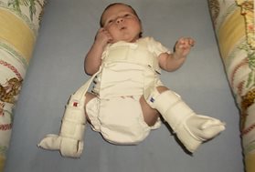
When infants need treatment for hip dysplasia, our first line of defense is a Pavlik harness. The harness is generally worn for 23 hours per day for approximately six weeks, but it is removable for bathing. The harness keeps the legs flexed and rotated in the right position for normal development of the hip joint.  After treatment with a Pavlik harness, we use physical exams, ultrasound and X-rays to monitor growth and confirm the hip joint is developing properly.  Most children require no further orthopedic treatment after wearing a Pavlik harness.

Learn more about our treatment and research in DDH and other conditions affecting newborns.

1Yang S, Zusman N, Lieberman E, et al. Developmental Dysplasia of the Hip. Pediatrics. 2019;143(1):e20181147
Share Your Story: Andi’s Life Changing Journey

Share Your Story: Andi’s Life Changing Journey

Meet Andi, a patient who is treated by our spine and hip experts. Learn more about her journey below.

Blog written by Andi’s mom, Tera, of McComb, Mississippi. 

Andi’s life changing journey started the moment she was born. At birth, we were told something was severely wrong with her hips and we would need to double diaper her to keep her hips spread apart. Nobody actually went into details with us until later at our post-delivery follow-up when Andi was three days old. At that time, our pediatrician told us he had never heard or felt a clunk in hips the way Andi’s hips were reacting to the hip check. Our pediatrician told us he thought she had hip dysplasia and we would need to meet with an orthopedic doctor.

We were initially referred to Children’s Hospital of New Orleans and Andi was just 1 week old when we had our first appointment with the orthopedic doctor. She was officially diagnosed with bilateral hip dysplasia and was put into a Pavlik harness, which was to be worn 24/7 for three months. We did harness adjustments every couple of weeks and after three months, the X-rays showed that the left hip responded to treatment, but the right hip did not. Our doctor, at the time, decided to try the Rhino abduction brace. It was then that we noticed that Andi was in a lot of pain. Hip dysplasia is generally not painful, but for her it was very painful.

When you touched her right leg or made any hip movement, her entire spine curved like a ‘C’ and she screamed in pain. She had X-rays done on her spine, but this did not give us any answers. We were told her spinal curvature (32 degrees) was not severe enough to be true scoliosis.
 
My husband and I began to research our options and that is when we found Texas Scottish Rite Hospital for Children. We live eight hours away and did not even hesitate about travelling to Dallas for a second opinion. The appointment process was quick, easy and we were able to get something scheduled right away.

Our first appointment was wonderful. 

Andi had Dr. Ramo and the staff scratching their heads, but they never gave up on her. They saw how much pain she was in and were able to pick up on developmental delays that nobody else had mentioned before. X-rays showed her right hip was still out of socket, her spinal curvature was now 42 degrees and the pain she was experiencing was being caused by inflammation.

Dr. Ramo suggested we allow Andi to continue to grow and develop and the plan was to repeat her scans in a couple of months. Time passed and we made another trip to Dallas. Her scans showed that her spinal curvature had since progressed to 54 degrees and Dr.Ramo decided to focus on her spine before we continued with further hip treatment. She was then placed in a Mehta cast – this process was rather simple, and the hospital staff made it easy.
 

From the moment we walked in the doors of the hospital to when we were discharged, our family felt comfortable, safe, loved and we knew our daughter was receiving the best care possible.

 
Fast forward a year later and Andi’s last Mehta cast was removed. Her spinal curvature had improved to 28 degrees and we were over joyed. She continues with nighttime bracing for a few months and then decided it was time to fix her hip.

Andi underwent a pelvic Osteomoy of the right hip and was placed in a unique spica/Mehta combination cast. She was in the combo cast for 13 weeks and at the end of this treatment, her hip looked wonderful! She did lose a little correction in her spine, but Dr. Ramo knew this would improve once she was able to wear a brace. She wore a rhino abduction brace for a month and then we continued with nighttime bracing.
 
Andi has been such a trooper throughout this entire process. For her, this is her normal nighttime routine – take a bath and then put on her brace. We had a follow-up appointment in December 2018 and her spinal curvature is now just 16 degrees. We will continue with her nighttime brace until our follow-up this summer and hopefully then, she will become an observation patient.

Everyone at Scottish Rite Hospital – from the registration staff, to the volunteers, nurse Marviel, Dr. Ramo and the cafeteria staff – they are all amazing. So kind, helpful, caring and welcoming! All of the hospital volunteers are always so giving and constantly put smiles on people’s faces.

This hospital made our struggles bearable.

Additional information on Developmental Dysplasia of the Hip

DO YOU HAVE A STORY? WE WANT TO HEAR IT! SHARE YOUR STORY WITH US.