Content included below was presented at the 2021 Pediatric Orthopedic Education Symposium by pediatric orthopedic surgeon William Z. Morris, M.D.
You can watch the full lecture and download this summary.
In hip dysplasia, the acetabulum (or hip socket) is shallow and doesn’t adequately cover the femoral head. Developmental dysplasia of the hip (DDH) occurs in approximately 1% of newborn children, and it is associated with four risk factors:
- Female
- Firstborn
- Feet first (breech)
- Family history
Hip dysplasia is relatively more commonly diagnosed in skeletally mature adolescents, affecting around 3% to 5% of the asymptomatic population. Cross-sectional studies have shown that female sex and a family history of dysplasia remain risk factors in adolescents.
There has been growing attention to the treatment of hip dysplasia as there is an association between hip dysplasia and the development of early osteoarthritis. In 1939, Gunnar Wiberg first described hip dysplasia and objectively measured it using what is now called the lateral center edge angle, to describe how well the socket (acetabulum) covers the ball (femoral head). On an AP (anterior posterior) pelvis X-ray, the angle is created by a vertical line through the center of the femoral head and a line from the center of the femoral head to the lateral border of the acetabular fossa. A larger angle reflects greater hip coverage, and a smaller angle reflects less coverage, commonly seen in a dysplastic hip.
Wiberg followed patients with dysplasia for up to 30 years and found that all of the patients with hip dysplasia eventually developed osteoarthritis. The smaller their center edge angle was (reflecting greater hip dysplasia), the faster they developed osteoarthritis.
Development of Osteoarthritis in Dysplastic Hips
The development of early osteoarthritis is suspected to occur due to a couple mechanical reasons. The most important factor is that:
Pressure = Force / Unit Area
In the hip, pressure on the joint surfaces depends on the total surface area of the femoral head in contact with the socket. A well-covered femoral head distributes weight-bearing forces across a larger surface area, reducing the pressure on each unit of cartilage. In contrast, a dysplastic acetabulum offers less surface area which increases the pressure on the cartilage contributing to earlier hip degeneration. In addition to the smaller weightbearing surface, the acetabulum is also more obliquely oriented. Therefore, compressive forces are less and shearing forces are greatly increased. This increased shear force may also contribute to cartilage degeneration.
Symptoms of Adolescent Hip Dysplasia
There are many different causes of hip pain in an adolescent patient and combining clues from the history and physical exam is essential to determine the underlying problem. The location of a patient’s pain can help determine the underlying etiology. Intra-articular pain of the hip usually presents as anterior groin pain, often due to a cartilage injury, a tear in the labrum (the ring of cartilage at the periphery of the socket) or inflammation of one of the hip flexors called the Iliopsoas tendon. Lateral hip pain that locates over the greater trochanter (the bony prominence on the lateral aspect of the thigh) probably reflects trochanteric bursitis or inflammation of the bursa overlying that region. Pain or soreness after activity above the greater trochanter where hip abductors like the gluteus medius are located may signal hip abductor fatigue, which is common with hip dysplasia. Pain over the iliac crest where the abdominal musculature attaches could reflect some inflammation of that apophysis, of the anterior superior iliac spine (ASIS) where the sartorius attaches, or of the anterior inferior iliac spine (AIIS) where the rectus tendon attaches.
Additional Factors to Consider when Discussing Hip Joint Symptoms
- Pain
- Duration
- Aggravating factors
- Squatting, stairs, low chairs
- Alleviating factors
- Mechanical symptoms
- Locking, popping, catching, snapping
- Neurological symptoms (which may suggest spinal pathology)
- Radiating pain, paresthesias
Physical Exam
- Pain Assessment
- Palpation can reproduce symptoms and help to localize the pain.
- Strength Testing
- Range of Motion
- Limited internal rotation (IR) can indicate hip impingement or a more acute concern, slipped capital femoral epiphysis (SCFE)
Special Tests
Straight Leg Raise Test
A straight leg raise is a passive test that helps to distinguish between hip and spine pathology and is performed by flexing the hip with an extended knee in a supine position. If this maneuver reproduces the patient’s pain that radiates distally, the problem may be related to nerve compression in the spine rather than a problem in the hip.
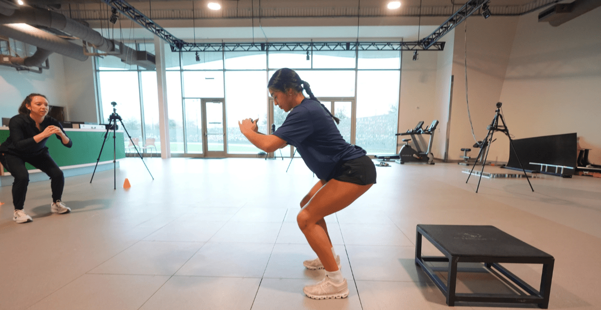
Trendelenburg Test
The hip abductors (e.g. gluteus medius) are typically weaker when patients come in with pain, so it can be targeted in physical therapy. The Trendelenburg sign is a quick physical examination used to assess for hip abductor weakness.
The patient stands on one leg (stance leg) and bends the other knee about 90°. Observe for evidence of hip abductor (i.e. gluteus medius) weakness which includes:
- Pelvis drop contralateral to the stance leg.
- Trunk lean/shift toward the stance leg.
Apprehension Test
Another test to further evaluate for dysplasia is the apprehension test:
- The patient lays in a lateral position
- Abduct the patient’s leg 30° away from midline
- Flex the patient’s knee 90°
- Gradually extend the hip
Patients who have an anterior uncovering of the socket from dysplasia will feel pain or a sensation of apprehension, which suggests that there may be some instability or dysplasia.
Femoroacetabular Impingement Test
Hip impingement should also be tested:
- Flex the hip to 90°
- Abduct the hip, bringing it towards midline
- Internally rotate the hip
This test attempts to reproduce hip impingement where the femoral head or neck collides against the socket. If this causes pain, there may be a cartilage injury such as a labral tear. An MRI is recommended to evaluate intra-articular soft tissues.
Slipped Capital Femoral Epiphysis – A condition not to be missed
Slipped capital femoral epiphysis (SCFE) is an adolescent disorder in which the growth plate is damaged and the femoral head epiphysis moves, or slips, with respect to the rest of the femur. Diagnosis in a timely manner is essential to prevent further injury to the hip.
Consider SCFE if these signs are present:
- limp
- walk with their foot externally rotated
- have limited range of motion, especially with internal rotation
Obligate external rotation is nearly pathognomonic for slipped capital femoral epiphysis, so if the patient can’t flex their hip straight up without turning their leg into an externally rotated position to accommodate further flexion, an anterior-posterior and frog pelvis film is recommended to ensure slipped capital femoral epiphysis hasn’t been missed. Immediate non-weight bearing with a wheelchair and urgent referral to pediatric orthopedics or an emergency room is recommended for this condition.
Radiographic Evaluation
A radiographic evaluation is important for a definitive dysplasia diagnosis. The AP pelvis film is a workhorse tool for the evaluation of hip coverage. It is taken standing to allow assessment of the patient’s hip coverage in their functional position using the lateral center edge angle (LCEA). A hip with an LCEA less than 25° is considered dysplastic.
Imaging also allows an assessment of the inclination of the socket. By drawing a line between the medial and lateral edges of the roof of the socket and measuring the angle between that line and a horizontal line, physicians can determine the acetabular inclination. The more inclined the socket is, the more dysplastic.
Treatments
Treatment for dysplasia begins with nonoperative options, which include:
- Physical therapy
- Activity modifications
- Non-steroidal anti-inflammatory drugs (NSAID)
When nonoperative treatments don’t work, and patients continue to have radiographic dysplasia and pain, including abductor fatigue pain above the greater trochanter or anterior intra-articular groin pain, they are treated surgically with a periacetabular osteotomy (PAO). This specialized procedure is done in patients who are approaching skeletal maturity or are already skeletally mature. Several cuts are made around the socket of the acetabulum to mobilize the socket. The socket is then reoriented to better cover the femoral head.
Outcomes
For patients with symptomatic hip dysplasia, the PAO has been shown to be successful in improving patients’ function, getting them back to their activities/sports, and preventing early hip replacement. In patients with hip pain and clinical or radiographic evidence of acetabular dysplasia, please consider a referral to Scottish Rite for Children for discussion of the condition and shared decision-making in the plan for management.
Check out our latest resources for medical professionals