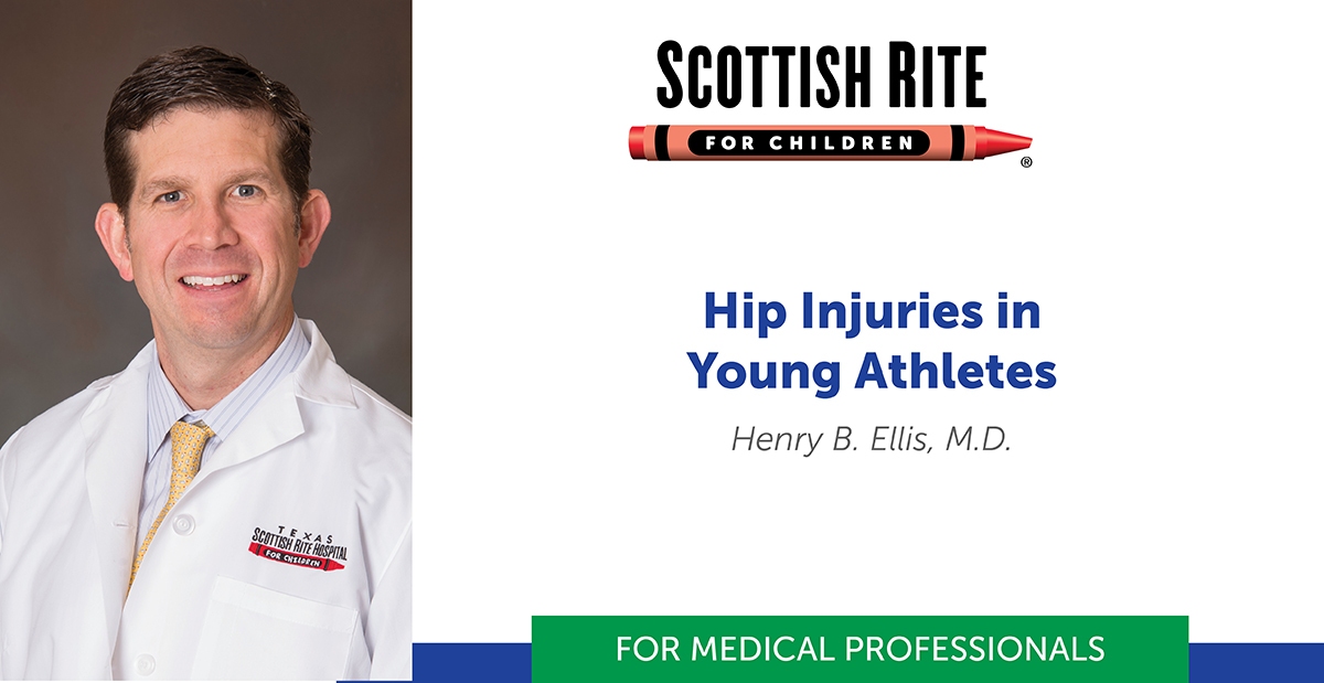
Pediatric orthopedic surgeon and associate director of clinical research, Henry B. Ellis, M.D., presented this as part of Coffee, Kids and Sports Medicine education series.
Watch the full lecture
Print the PDF
Ellis provided a detailed discussion of the history and physical exam of young athletes with hip complaints to distinguish between common and less common hip conditions. Young athletes require a different approach than an adult athlete. Numerous conditions present more often, or only, in a younger patient compared to an adult. These include slipped capital femoral epiphysis (SCFE), adolescent hip dysplasia, epiphyseal dysplasia, apophysitis, stress fractures and more.
The ball and socket joint allows motion in all planes. For some young athletes, the soft tissue is particularly flexible which can increase the risk for injuries. A review of the anterior-posterior (AP) pelvis X-ray in a growing child provides an excellent overview of the pertinent anatomy in the growing pelvis and hips. There are physes, growth centers, that are present early and active through adolescence. Pelvis and hip growth centers include:
- Acetabular physis (triradiate cartilage)
- Proximal femoral physis
- Greater trochanter apophysis
- Ischial tuberosity
- Anterior superior iliac spine
Five Key Tips for Evaluating the Youth Athlete’s Hip
- History can help focus the exam.
- Always examine both sides.
- Adequately expose the area of interest while maintaining modesty.
- Look beyond the hip.
- Consider chaperone or an assistant in the room with hip exam.
Ellis says his detailed hip exam will last about 15-20 minutes. To provide an overview, he demonstrated a “three-minute hip exam” before he provided a detailed explanation of each step discussing associated conditions with each step.
Tests for recognizing signs of concerning conditions:
- Passive hip flexion that causes obligate (automatic) external rotation is indicative of SCFE and requires a prompt referral to minimize sequelae.
- Dial test/passive circumduction with the hip joint relaxed. The provocation of pain indicates intra-articular problems such as synovitis or infection.
- Hip flexion (90+ degrees) with adduction and internal rotation that causes pain is a sensitive screening tool for labral pathology.
- Hip apprehension sign is positive when hip abduction and external rotation in side-lying causes apprehension and indicates a need for additional assessment for hip dysplasia.
In conclusion, Ellis provided some take-home messages for the audience.
- A good clinical exam will often lead you to the diagnosis.
- AP and frog pelvis X-ray is appropriate to evaluate for hip problems.
- 80% of hip injuries are soft tissue strains that can be treated with rest, early range of motion and gradual return to sports when pain improves.
- Some hip conditions require a MR arthrogram, so avoid an MRI of the hip until evaluated by a specialist, unless a stress fracture or other concerning diagnosis is suspected.
Ellis never disappoints an audience. As the first event after a break from our livestream events, we received these wonderful comments from attendees:
- “So great to be here again!”
- “Thank you for a well put together and thorough presentation. Also, I appreciate the handouts.”
Check out our latest on-demand lectures available for medical professionals.


