In approaching the treatment of a clubfoot, it is essential for families and providers to understand the presence of these congenital differences because our method of treating clubfoot cannot alter any of them. We can’t change the shape of the bones. We can’t improve the elasticity of the soft tissues. We can’t change the way the blood vessels move into the foot, and we can’t change the power with which certain muscles fire. Therefore, the goal of clubfoot treatment isn’t to create a normal foot. Rather, the goal of treatment is to take a structurally abnormal foot, which is also in an abnormal position, and through a series of casts applied weekly, slowly convert into an abnormal foot structurally that is ultimately in a near normal or completely normal position. Also, this method limits the need for surgery, which creates scarring and stiffness, thereby preserving as much motion of the foot as possible.
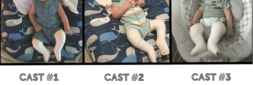
The Ponseti method of treatment is the gold standard of care worldwide for clubfoot deformity. This involves weekly serial casting of the involved foot or feet, with each cast applied in a manner to correct a different component of the clubfoot deformity. These casts are very effective at correcting every component of the clubfoot deformity with great success, save for the contracture of the Achilles tendon, which drives an oftentimes rigid equinus (plantarflexion) deformity of the ankle. Approximately 90% of clubfeet require a percutaneous transection of the Achilles tendon. This is performed in the clinic with topical anesthesia in children aged less than 3 months to overcome this component deformity. Following the heel cord tenotomy, the ankle can be acutely dorsiflexed to at least 10 to 15 degrees, and that position is maintained in a final cast that is kept in place for three weeks.
On average, it takes about four weeks of weekly casting to correct the component deformities of the cavus, hindfoot varus, and metatarsus adductus. That brings the foot from what is essentially an upside down and turned inward position to a right-side-up and turned-out position. At that point, if the ankle remains in equinus, this deformity is corrected with a heel cord tenotomy and a final three-week cast, resulting in a total time to achieve full correction of about seven weeks. As we ideally begin treatment within one or two weeks of life, most deformities are corrected by or before 2 months of age.
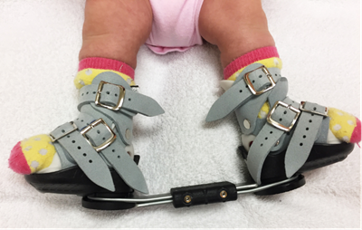
Though bracing protocols vary to some degree, our protocol mirrors that agreed upon at the International Clubfoot Congress and consists of brace use for 23 hours a day until the child begins to pull to stand and 12 hours a night thereafter until age 4. The braces do not interfere with the achievement of developmental milestones, such as sitting up, rolling over, crawling or standing. The transition from full-time to part-time use is based entirely on the fact that once a child is able to pull to a standing position, they will soon spend more time on their feet. At that time, their own body weight will help to keep their feet flat on the ground. Until that time, babies spend most of their time lying down and sitting, during which the feet are held in a relaxed position. That can result in tightening of the heels cords and a recurrence of equinus.
Bracing is most difficult on parents for the first two weeks after the child comes out of their post-tenotomy cast. After seven weeks of casting, having anything other than cast padding surrounding their feet feels different and can cause fussiness. Parents are counseled extensively about this so they can anticipate and develop strategies to keep the braces in place despite frustration on the part of the child.
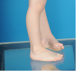
Unfortunately, even when perfect correction is achieved with casting, and parents are compliant with brace use, a percentage of these deformities still recur and require additional treatment due to the structural congenital differences present. Research has demonstrated that with parental bracing compliance, about 80% of clubfoot patients never need any additional treatment. However, despite everyone’s best efforts, 20% of children still need some additional treatment due to recurrence. These recurrent deformities are most commonly the result of an imbalanced muscle pull across the foot due to a relatively overpowered tibialis anterior tendon. When relatively overpowered, this muscle, due to its insertion along the medial aspect of the foot, supinates the foot during dorsiflexion. That results in the touchdown of the lateral border of the foot during the initial stance phase of gait. This supination, in turn, slowly drives the foot inward and into further supination with weightbearing. Such recurrences are easily managed with a brief period of repeat casting to correct any relapsed deformities. That’s then followed by a transfer of the offending tendon to the dorsum of the foot to remove that supination moment during dorsiflexion.
With an experienced team of providers and parental dedication to brace use, these initially dramatic foot deformities can be corrected beautifully and without any long-term deficits or disabilities. However, it is important to educate parents early regarding certain differences in the foot or feet following treatment. A clubfoot is always smaller than a normal foot, sometimes half to a full shoe size smaller than an unaffected contralateral foot. In addition, the calf musculature of children with clubfoot tends to be smaller, though this size difference does not appear to affect strength. These differences are strictly cosmetic and do not alter function. The goal of clubfoot treatment is to allow a foot or feet to sit flat to the ground, have as much flexibility as possible, allow the child to wear any shoes they want, play any sports they want, have any job they want, and do whatever they want with the foot. In achieving this, their corrected clubfoot will function no differently than a normal foot and will never limit a child in any way in any activity they desire.
Download the PDF.
Dr. Anthony I. Riccio is a pediatric orthopedic surgeon and the Director of the Center for Excellence in Foot at Scottish Rite for Children.