This article was recently published in the Pediatric Society of Greater Dallas newsletter. Committed to improving orthopedics care of pediatric patients in all settings, Scottish Rite for Children specialists are regular contributors to this publication for local pediatricians.
What are Juvenile Bunions?
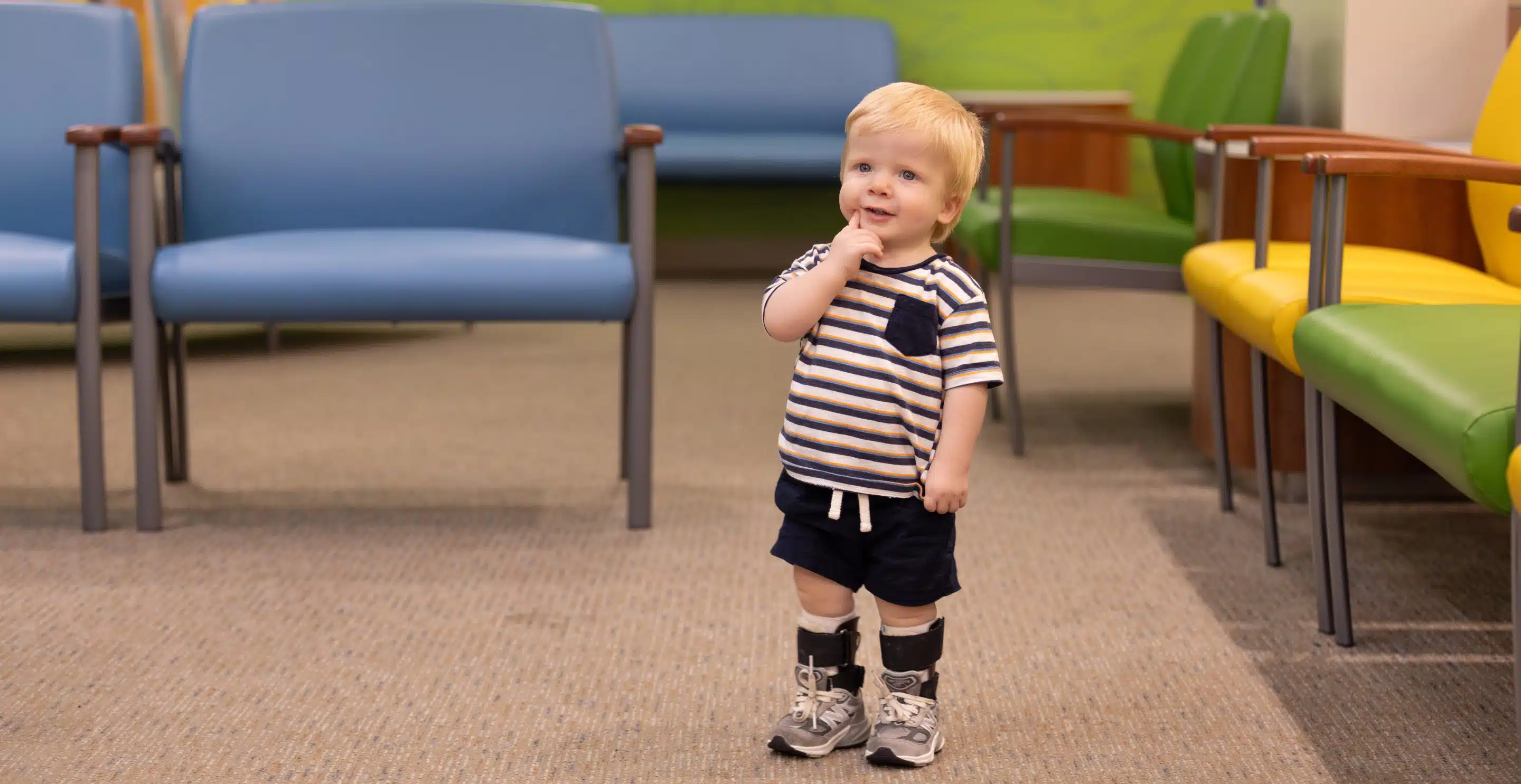
Juvenile bunions, a deformity of the foot, are a common cause of frustration for patients, their parents and medical providers. Fortunately, the vast majority of bunion deformities in children and adolescents are asymptomatic and therefore require no intervention aside from parental reassurance. For those deformities that cause difficulty with shoe wear and pain refractory to conservative treatment modalities, surgical correction is a reasonable option. It cannot be overstated, however, that appropriate surgical management mandates an in-depth understanding of the component deformities to avoid the historically high rate of recurrence and failure following operative treatment.
How Do Adult Juvenile Bunions Differ From Adult Bunions?
Clinically, adult and juvenile bunions appear very similar with lateral deviation of the great toe and a medial eminence at the metatarsophalangeal (MTP) joint resulting from uncoverage of the medial metatarsal head. These are, however, very different deformities. Adult bunions are
most commonly acquired deformities from tight shoe wear which leads to attenuation of the medial sided soft tissues at the MTP joint, lateral deviation of the hallux and usually pronation of the toe as well.
What Causes Bunions?
The cause of juvenile bunions is neither completely understood nor agreed upon. Hypermobility of the first ray, an associated flatfoot and obliquity of the lateral cuneiform have all been postulated as causative factors. However, the deformity is most likely congenital and results from external orientation of the articular surface of the metatarsal head.
How Do Bunions Appear in Radiographs?
Radiographically, both adult and juvenile bunions are characterized by angulation between the first metatarsal and the proximal phalanx (hallux valgus angle or HVA) as well as an increase in the angle subtended by the axes of the first and second metatarsals (intermetatarsal angle or IMA). The distinguishing characteristic of the juvenile deformity is a more lateral orientation of the distal metatarsal articular surface. Whereas in adult bunions, the articular surface is usually perpendicular to the metatarsal shaft. In younger patients, the angle between the shaft and the articular surface (distal metatarsal articular angle or DMAA) is elevated. This results in lateral deviation of the toe with maintained congruency of the MTP joint. In contrast, adult bunions usually demonstrate varying degrees of joint incongruency between the base of the proximal phalanx and the metatarsal head.
Can Bunions Be Prevented?
Because juvenile bunions are congenitally acquired, there are no means by which the deformity can be prevented. Moreover, once noticed, no well-accepted means of stopping deformity progression are available. Commercially available toe spacers, bunion straps and physical therapy cannot alter the abnormal anatomy of the distal metatarsal articular surface and therefore have no role in the management of this clinical entity. Straps and spacers that force the great toe more medially and the give the outward appearance of a straighter hallux do so by creating incongruity at the MTP joint which can alter range of motion and oftentimes cause discomfort.
What Shoes and Pads Help with Juvenile Bunions?
As mentioned above, in the absence of pain or difficulty with shoe wear, parental reassurance is the best course of action. In the presence of symptomatic deformities, nonoperative measures are all that is usually required to keep children active and happy. Shoe wear modifications are the simplest and oftentimes most effective course of action. Adolescents and their parents should be encouraged to find a well cushioned stability shoe with a wide toe box and flexible uppers. For those children required to wear leather shoes, boots or cleats due to school uniform requirements or recreational desires, most cobblers can create extra space in the region of the medial eminence using a “ball and ring” shoe stretcher. Should shoe modifications fail to provide complete relief, silicone bunion pads placed over the medial eminence can also be helpful. In children with flexible flatfoot deformities and symptomatic bunions, using a soft shoe insert to elevate the medial half of the heel and support the arch can offload any plantar-medial pressure by elevating the medial column of the foot.
Is Surgery Necessary for Bunions?
When prolonged conservative treatments fail to provide relief, surgical intervention is a reasonable option. Though recommendations vary with regard to the timing of surgery, due to historically poor results following the surgical treatment of juvenile bunions, many advocate to delay surgery until adolescents are within one or two years of skeletal maturity.
Females typically reach skeletal maturity at age 14 while males reach skeletal maturity at age 16. Better understanding of the congenital nature of the juvenile bunion and the importance of correcting the DMAA during operative treatment will likely lead to a dramatic reduction in recurrence and surgical failure, ultimately driving down the age at which operative intervention can be successfully undertaken.
Who is Qualified to Perform Surgery to Correct Juvenile Bunions?
As such, surgery should be performed only by those with a clear understanding of this specific deformity such as a fellowship-trained pediatric orthopedic surgeon or a fellowship-trained orthopedic foot and ankle specialist who is comfortable caring for pediatric patients.
Anthony I. Riccio, M.D., is a pediatric orthopedic surgeon caring for children and adolescents at Scottish Rite for Children. He is the director for the Center for Excellence for Foot.