Understanding Hip Impingement in Teens: How it Happens and How to Prevent It

Understanding Hip Impingement in Teens: How it Happens and How to Prevent It

Also commonly referred to as hip impingement, femoroacetabular impingement is a painful condition that occurs in the hips of adolescents and young adults. Two bones fit together to make up this “ball and socket” joint including the head of the femur (ball), which is part of the thigh bone, and the acetabulum (socket), which is part of the pelvis.

Impingement, or pinching, causes pain when the bones in the hip joint pinch the labrum, the soft tissue on the perimeter/edges of the acetabulum.
There are three types of FAI:

  • Cam impingement occurs when the shape of the femoral head or ball is abnormal.
  • Pincer impingement occurs when the shape of the acetabulum or socket is abnormal.
  • Combined impingement occurs when both the ball and the socket are abnormal.

Pediatric orthopedic surgeon Henry B. Ellis, M.D., says, “Repetitive activities make changes in the joints. In the hip, either the soft tissues become damaged, the bone actually changes its shape or both of these occur.” The reason for abnormal bone shape is not known. It may occur during development or may be in response to activity.

Symptoms of hip impingement are more likely to occur in those who perform:

  • Repetitive maximal flexion (bending) of the hip, such as deep squatting or high kicking.
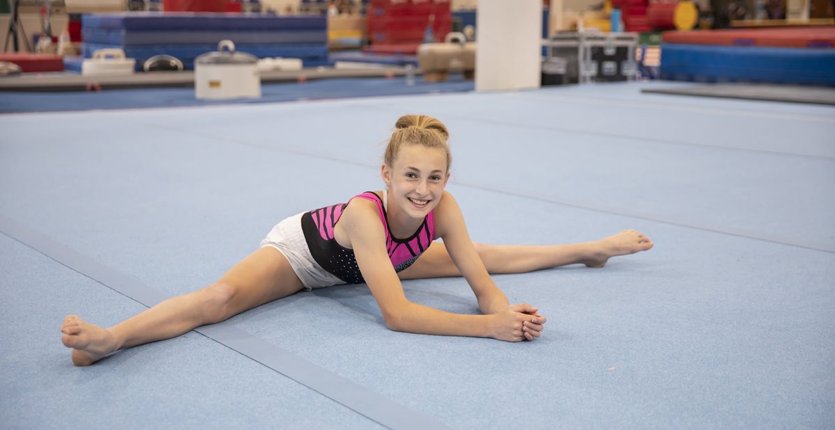
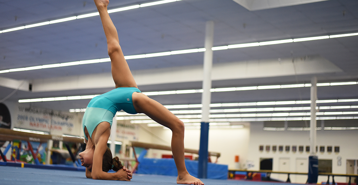
  • Repetitive movements in activities, such as running, dance, gymnastics and hockey.

What are the symptoms of femoroacetabular impingement?

  • Pain in the hip or groin, typically in the front.
  • Tenderness and/or swelling of the hip or groin area.
  • Stiffness or pain after sitting for long periods of time.
  • Aching or pain that worsens with certain activities.

How is it diagnosed?
A thorough history and physical examination are used to diagnose a hip impingement. In most cases, X-rays are used to further assess the shape and fit of the bones. If symptoms do not improve or worsen, additional imaging such as an MRI or MR arthrogram may be recommended to further evaluate the soft tissue, the acetabular labrum. An MR arthrogram uses MRI, fluoroscopy and sometimes an injected medication to show the structures inside the joint.
 
How is it treated?
Treatment depends upon the severity of the condition and typically begins with a nonoperative approach which typically includes resting from activities that cause pain or changing to activities that do not. Other treatment options include physical therapy, joint injections or arthroscopic surgery may be required.
 
In a recently published article “Risk Factors for Suboptimal Outcome of FAI Surgery in the Adolescent Patient”*, Ellis and others reported findings after reviewing 126 hips (114 patients) under the age of 18 who were being treated for symptomatic FAI. This work helps Ellis and his colleagues around the country provide better counseling to patients considering surgery for FAI.
 
Early recognition and treatment are important because hip impingement has been shown to be a risk factor for early development of osteoarthritis of the hip.
 
How can hip impingement be prevented?
Overuse injuries like hip impingement and FAI occur with a high volume of training, repetition of certain movements and early specialization in a sport.
 
“Hip impingement in a growing child is bad news. We need to help them monitor and modify their volume of repetitive activities to prevent the condition from worsening, or even better, developing.”

  • Henry B. Ellis

These suggestions can help to prevent FAI and other similar overuse conditions:

  • Avoid sports specialization and play multiple sports throughout high school.
  • Emphasize moderation with load and training.
  • Encourage free play and lifetime sports like cycling and hiking.
  • Avoid year-round participation and encourage weekly and seasonal rest from activities requiring repetitive maximal flexion of the hip.
  • Perform proper warm-up and conditioning for all activities.                                                           

Learn more from Ellis about Hip Injuries in Young Athletes.
 
*Yen, Y. M., Kim, Y. J., Ellis, H. B., Sink, E. L., Millis, M. B., Zaltz, I., Sankar, W. N., Clohisy, J. C., Nepple, J. J., & ANCHOR Group (2024). Risk Factors for Suboptimal Outcome of FAI Surgery in the Adolescent Patient. Journal of pediatric orthopedics44(3), 141–146.

Keeping Up with the Count: Hip Health in Dancers

Keeping Up with the Count: Hip Health in Dancers

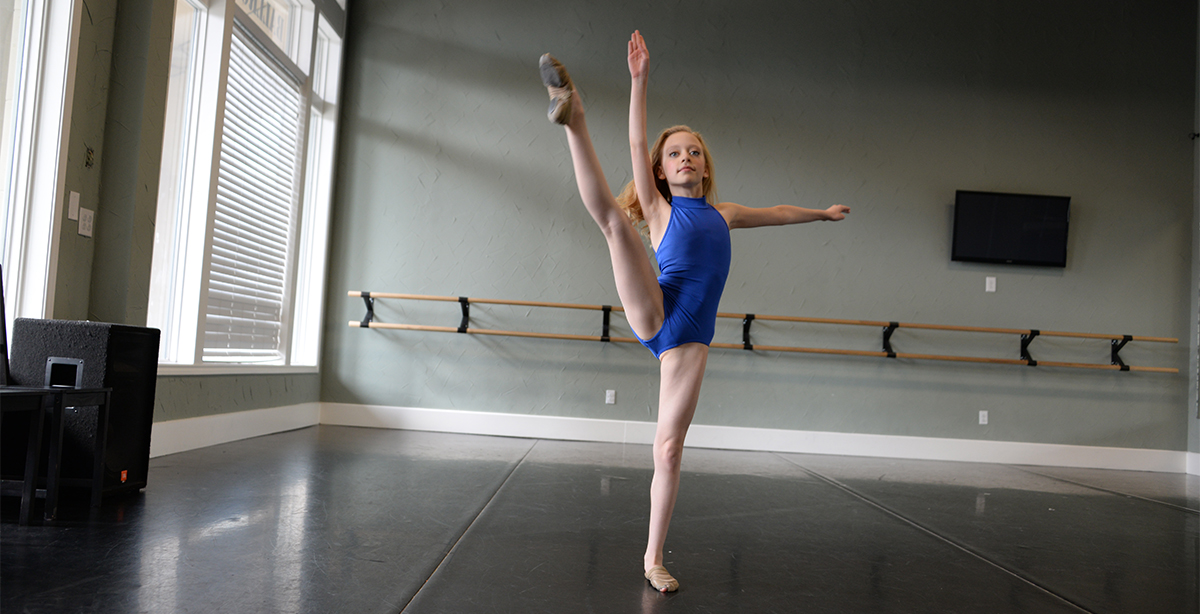
Dancers and other performing artists place demands on their hips that are unlike those of other athletes. Movements push the range of motion of their hips to extreme ends from an early age. They must have the flexibility for turnouts, leaps, or grand battements, and also absorb dramatic forces from leaps and jumps. All of this while maintaining impressive limb control and stability of their support and gesture limbs over sustained periods.

Key Considerations for Hip Health for Dancers

  1. Mobility in the hip and surrounding muscles.
  2. Balance and stability in the pelvis and core.
  3. Managing training volume.

Hip Mobility
Turnout and extreme ranges of motion during dancing and other performing arts often require more than “normal” hip mobility. For some, the end of the thigh bone in the hip is naturally in a position of retroversion, which allows for the extreme external rotation needed for turnouts. Others have soft tissue laxity, also called joint hypermobility, that predisposes them to successfully achieve the extremes in rotation, extension, flexion, and abduction (out to the side, such as a la seconde) required for their art form. Dancers with natural hypermobility may be more likely to continue, whereas others may “self-select” out of the sport.

Dancers that are not born with these factors may acquire laxity in the joint with many years of training and aggressive stretching of the muscles and soft tissue that make up the hip capsule.

Proper supervision and a comprehensive program are necessary to ensure the stretching does not cause hypermobility in the lower spine. Additionally, extreme motions may cause damage to the labrum, a soft tissue rim that stabilizes the hip in the socket. Therefore, prompt response to signs of pain with mobility should be addressed to avoid damage to the soft tissue, and ultimately the bones in the hip.

Pro Tip: When the core stabilizer muscles don’t support the lower spine, the hip muscles, including the flexors or hamstrings, are forced to provide support. This protective tightness is an undesirable compensation and can be corrected by doing core stabilization exercises.

Pelvic and Core Stability
Mobility of the hip and leg is dependent on having a stable platform. Core stability means abdominal strengthening to many, but there are deeper muscles that must be considered, including:

  • Gluteal muscles – deep hip rotators that help to maintain active turnout and appropriate knee alignment in the posture leg with grand plies and more.
    • Gluteus maximus (hip rotation and extension)
    • Gluteus medius (hip rotation and abduction)
  • Transverse abdominis – deep abdominal muscles

Imbalances and weakness of these muscles cause stress on other joint tissues, including the capsule, labrum, and ligaments. Stretching or stressing beyond their limits can cause pain and injuries in those non-muscular tissues, which then shifts more demand to the muscles around the hip to provide extra support at end ranges of motion. The body then uses other muscles like the hip flexors to stabilize the hip and support a high volume of hip flexion with a turned-out leg, as seen in dance.

A consequence of this demand or overuse of a muscle is muscle tendinitis, the inflammation of the tendon part of a muscle. This condition worsens when there are sudden spikes in the frequency or duration of training, particularly when there is inadequate support from the core to control the pelvis during repeated hip flexion movements.

Stability Exercises:

Abdominal Hollowing Technique
To prevent this chain of compensation, a dancer can learn how to activate the transverse abdominis, the deep abdominal muscles. These muscles help to create a stable base prior to limb movement. Activation of these muscles is described as a “hollowing” technique as the belly button is pulled inward toward the spine. This contrasts with a “bracing” technique that activates the superficial abdominals.

Pro Tip:
A dancer should be able to do an active straight leg raise without any arching of the lower back during the movement. For added abdominal/core muscle activation, use a band for a pull-down during the straight leg raises. This prepares a dancer for flexion associated with high kicks and grand battements without anterior pelvic tilt.

Gluteal Medius Strengthening

Example exercises:

  1. Single leg glute bridge
  2. Kneeling, side plank, hip abduction raises
  3. Clamshell side planks
  4. Side plank development

Pro Tip: It is important to learn to use the gluteus medius instead of the spinal muscles, called the quadratus lumborum, with abduction motions out to the side (a la seconde) or in the posture leg.

Manage Training Volume
Poor form and muscle fatigue can cause undesirable compensations with other soft tissues and muscles, and may lead to direct tissue injury in the joints or muscles. As overuse injuries worsen with time, performance suffers when the muscles are fatigued.

Pay attention to sudden increases in training duration or intensity, such as fall preparation for The Nutcracker, when added to typical training classes because it can leave a dancer vulnerable to injury. Dancers should take a day off one to two times each week for recovery. Proper rest can help prevent injuries, so you stay healthy throughout the season.

Hip Injuries in Young Athletes

Hip Injuries in Young Athletes

Pediatric orthopedic surgeon and associate director of clinical research, Henry B. Ellis, M.D., presented this as part of Coffee, Kids and Sports Medicine education series.

Watch the full lecture
Print the PDF

Ellis provided a detailed discussion of the history and physical exam of young athletes with hip complaints to distinguish between common and less common hip conditions. Young athletes require a different approach than an adult athlete. Numerous conditions present more often, or only, in a younger patient compared to an adult. These include slipped capital femoral epiphysis (SCFE), adolescent hip dysplasia, epiphyseal dysplasia, apophysitis, stress fractures and more.

The ball and socket joint allows motion in all planes. For some young athletes, the soft tissue is particularly flexible which can increase the risk for injuries. A review of the anterior-posterior (AP) pelvis X-ray in a growing child provides an excellent overview of the pertinent anatomy in the growing pelvis and hips. There are physes, growth centers, that are present early and active through adolescence. Pelvis and hip growth centers include:

  • Acetabular physis (triradiate cartilage)
  • Proximal femoral physis
  • Greater trochanter apophysis
  • Ischial tuberosity
  • Anterior superior iliac spine

Five Key Tips for Evaluating the Youth Athlete’s Hip

  • History can help focus the exam.
  • Always examine both sides.
  • Adequately expose the area of interest while maintaining modesty.
  • Look beyond the hip.
  • Consider chaperone or an assistant in the room with hip exam.

Ellis says his detailed hip exam will last about 15-20 minutes. To provide an overview, he demonstrated a “three-minute hip exam” before he provided a detailed explanation of each step discussing associated conditions with each step.

Tests for recognizing signs of concerning conditions: 

  • Passive hip flexion that causes obligate (automatic) external rotation is indicative of SCFE and requires a prompt referral to minimize sequelae.
  • Dial test/passive circumduction with the hip joint relaxed. The provocation of pain indicates intra-articular problems such as synovitis or infection.
  • Hip flexion (90+ degrees) with adduction and internal rotation that causes pain is a sensitive screening tool for labral pathology.
  • Hip apprehension sign is positive when hip abduction and external rotation in side-lying causes apprehension and indicates a need for additional assessment for hip dysplasia.

In conclusion, Ellis provided some take-home messages for the audience.

  • A good clinical exam will often lead you to the diagnosis.
  • AP and frog pelvis X-ray is appropriate to evaluate for hip problems.
  • 80% of hip injuries are soft tissue strains that can be treated with rest, early range of motion and gradual return to sports when pain improves.
  • Some hip conditions require a MR arthrogram, so avoid an MRI of the hip until evaluated by a specialist, unless a stress fracture or other concerning diagnosis is suspected.

Ellis never disappoints an audience. As the first event after a break from our livestream events, we received these wonderful comments from attendees:

  • “So great to be here again!”
  • “Thank you for a well put together and thorough presentation. Also, I appreciate the handouts.”

Check out our latest on-demand lectures available for medical professionals.

Apophysitis of the Hip or Pelvis

Apophysitis of the Hip or Pelvis

The professionalization of youth sports has led to an epidemic of overtraining. With that comes an increase in injuries caused by overuse. Unlike injuries from overuse in adults, like carpal tunnel syndrome, pediatric overuse injuries occur at areas of the bones called growth centers. These areas are vulnerable to injuries. Pediatric orthopedic surgeon Henry B. Ellis, M.D., says, “Rest and activity modification is crucial for these conditions.”
With an interest in studying and treating conditions in the hip in athletes, Ellis informs families that other treatments are unlikely to work if the aggravating activity continues. He encourages athletes to listen to their bodies and learn to properly stretch and to speak up if there is activity-related pain.

What is apophysitis of the hip or pelvis? 

Tenderness in specific bony areas of the hip and pelvis is called apophysitis. This typically occurs in adolescents ages 10- 19 who have “tight” hip and thigh muscles.

What causes apophysitis in hip or pelvis?

Muscles of the hip attach on the pelvis and upper leg bones. In growing children, several tendons attach to apophyses (growth plates). On the pelvis, these include the iliac crest apophysis and the ischial tuberosity apophysis and on the hip, the greater trochanter apophysis. These areas are made up of soft cells called cartilage. These weaker cells are at a higher risk of injury.

The most common cause of the pain is repeated pulling of the tendons on the apophysis causes apophysitis (painful inflammation). This commonly occurs during periods of rapid growth or increased activity. Overuse in these areas occur in activities such as dance, gymnastics and those that include running and sprinting.

What are the symptoms of apophysitis?

Apophysitis causes pain or tenderness at the muscle attachment that worsens with activities such as sports or running. Some experience swelling, others feel or hear a pop or snap.

How is it diagnosed?

A thorough history and physical examination are used to diagnose apophysitis. In some cases, X-rays may be ordered to evaluate the growth plate and rule out other issues.

How is it treated?

This is a self-limiting condition where rest is recommended, but kids may participate in activities that do not cause painful symptoms. Treatment includes modifying activities and providing comfort as needed. Learning to properly stretch and strengthen the muscles attached to the apophysis will reduce the tension.

A gradual return to sports is recommended when pain is improved. Symptoms will resolve with completion of growth in this area. In time, stronger bone cells replace the soft cartilage cells, but pain may come and go for months to years. If symptoms persist and cannot be managed with rest, it is important to see a medical provider with experience treating growth plate conditions in young athletes. Physical therapy may be recommended.

Physical therapy for apophysitis

Since the condition requires rest and removal of aggravating factors, more exercise is not an appropriate solution. After a formal evaluation, a physical therapist will provide a custom exercise plan to promote improvements in lengthening of tight muscles and tissues, strengthening of weak muscles and alignment of the body during movement and functional tasks. In many cases, core conditioning, strengthening of the abdominals and other trunk muscles, is a foundational component of treatment.

How can apophysitis of the hip/pelvis be prevented?

  • Apophysitis of the hip/pelvis may be difficult to prevent.
  • Proper warm-up and stretching exercises will reduce the stress on the apophysis.
  • Limit or vary physical activities to avoid overtraining and overuse.
  • Rest when sore or having pain.

Watch Ellis describe this condition in a series for pediatricians and primary care providers on our YouTube playlist for medical professionals.