Content included below was previously presented at the 2021 Pediatric Orthopedic Education Symposium by pediatric orthopedic surgeon Megan E. Johnson, M.D.
People hear the term scoliosis often, but they may not know what it means. Pediatric orthopedic surgeon Megan E. Johnson, M.D., walked through each phase of detecting and treating scoliosis in a recent lecture. This summary provides health care professionals with a succinct summary and language to navigate the steps and conversations with patients presenting with suspected scoliosis.
Watch the full lecture or download this summary
Defining scoliosis
Scoliosis is a structural lateral rotated curvature of the spine. For a condition to qualify as scoliosis, the Cobb angle, or the measurement of the degree of side-to-side spinal curvature, must be a minimum of 10 degrees. If the Cobb angle is less than 10 degrees, it is considered a spinal asymmetry, not scoliosis.
What are the types of scoliosis?
- Idiopathic scoliosis: The most common type of scoliosis. Idiopathic means that all other causes of scoliosis have been ruled out.
- Congenital scoliosis: When vertebral malformations cause a curvature of the spine. The vertebrae weren’t formed correctly or haven’t separated from the other surrounding vertebrae correctly.
- Neuromuscular and syndromic scoliosis: Occurs in patients with underlying neurologic disorders like cerebral palsy, spina bifida and other genetic conditions.
What age is scoliosis diagnosed?
Scoliosis may be diagnosed at any age, but earlier recognition often improves treatment options and outcomes. A patient’s age also helps to define the type of scoliosis that they may have.
- Infantile idiopathic scoliosis: patient is between 0 and 3 years old at the time of diagnosis.
- Juvenile idiopathic scoliosis: patient is between 4 and 9 years old at the time of diagnosis.
- Adolescent idiopathic scoliosis: patient is 10 years or older at the time of diagnosis.
The most common type is adolescent idiopathic scoliosis (AIS). The patient’s curve typically goes to the right and can include either the thoracic or lumbar spine or both.
What history and physical exam findings are important with scoliosis?
In order to evaluate patients, it is important to learn if patients have had back pain, headaches, other neurologic symptoms or a family history of scoliosis. For girls, it is also necessary to know their menstrual history to gauge where they are in their growth cycle.
The physical exam is focused on identifying asymmetries in static posture. This includes:
- Differences in shoulder height.
- Scapular asymmetry – scapulae are at different heights or one is more retracted.
- Pelvic obliquity – iliac crests are at different heights.
- Trunk shift – drawing an imaginary line from the patient’s head to their waist and seeing if the head is centered over their waist.
- Waist asymmetry – a visible bulge (typically on left) on the convex side of a lumbar curve, and crease on the concave side of the curve.
Special test
A very common test for scoliosis that most people are familiar with is the Adams forward bend test. Patients bend forward at the waist, and the examiner looks for signs of rotational deformities. Curves in the coronal plane cause rotation in the axial plane, which are visible in the Adams test. For example, a left midline lumbar prominence and the prominence of the right ribs are evident with a right thoracic curve.
Neurologic exam
A thorough neurologic exam assesses for asymmetries and changes in sensation, reflexes and motor function in the trunk and lower extremities. The patellar tendon, the Achilles tendon and the abdomen are tested to look for symmetry in reflexes. Being hyper-reflexive is fine when it is present on both sides. If there are reflexes on one side and not the other, it is an abnormal finding. Foot abnormalities, like a cavovarus foot or a significant flat foot on just one side, may indicate an underlying neurologic concern.
What radiologic imaging is used to diagnose scoliosis?
Plain film radiology (X-rays)
X-rays are essential to assess the patient’s scoliosis. Full-length X-rays of the spine, including the pelvis and the top parts of the hips and femurs, will give physicians all the information that they need to determine what the curve looks like, how big the curves are and how much growth the patient has left. Full-length X-rays are necessary for final diagnosis and treatment planning. Scottish Rite for Children uses advanced imaging technology called EOS, which utilizes a very-low-dose radiation for efficient and effective full-length images. To avoid unnecessarily repeating X-rays, images are not required for referrals for suspected scoliosis.
Advanced imaging
An MRI is indicated with these findings:
- Curve abnormalities like a left-sided curve, a back that is rounder than expected or an abnormal appearing curve.
- Short and sharp curves and kyphosis are red flags requiring further evaluation with an MRI.
- Abnormal neurologic exam or other neurologic symptoms, like daily headaches.
- Significant progression in the patient’s curve between follow-up appointments.
What factors are considered in planning treatment for scoliosis?
The goal of scoliosis treatment is to keep the spinal curve(s) as small as possible and prevent progression to surgery. The following are considered:
- Age of the patient.
- How skeletally mature they are.
- Size of the scoliosis curve(s).
Bracing
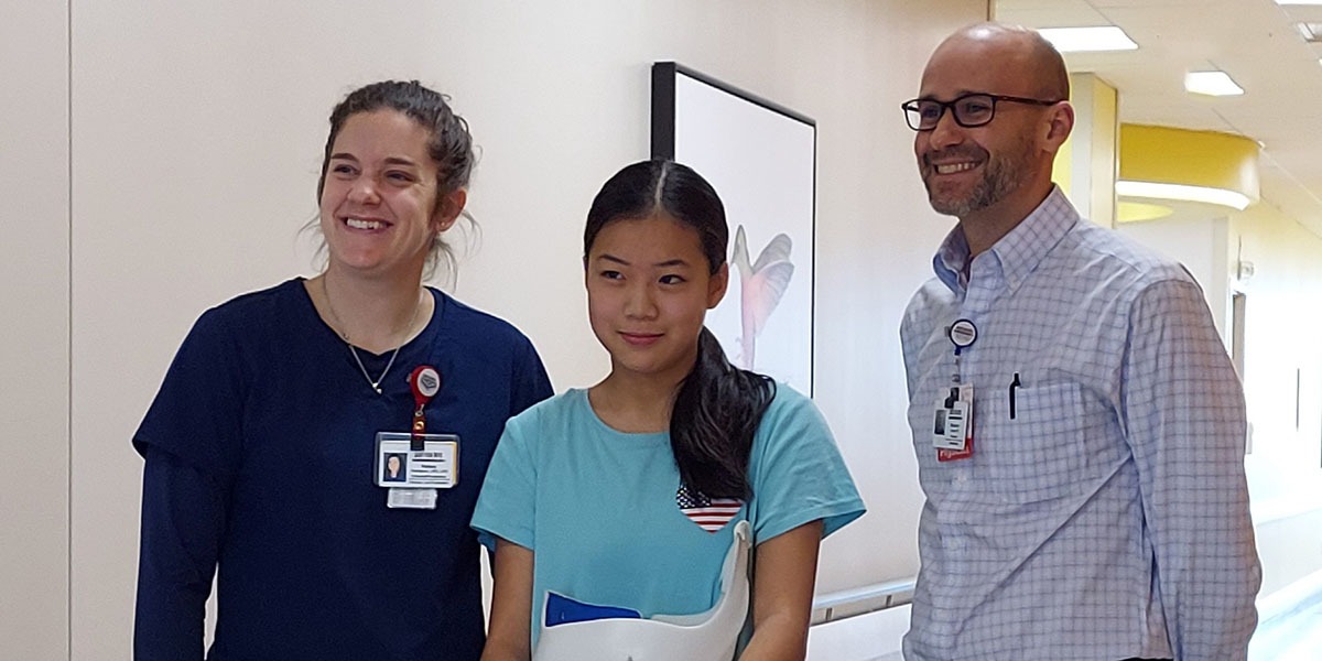
Bracing is recommended for patients with curves between 20-25 and 40 degrees if they have at least two years of growth remaining. Thoracic lumbar sacral orthosis (TLSO) braces are worn 18 to 20 hours a day and are typically used for curves in the thoracic spine or both the thoracic and lumbar spine. If the patient only has a lumbar curve that is flexible, nighttime bracing may be recommended.
Surgery
Surgery is recommended when the patient’s curve has a Cobb angle of 50 degrees or more to prevent the curve from progressing into adulthood. Surgery is generally not recommended until the patient is at least 10 years old because if the fusion is done too early, the growth of the patient’s spine can cause some secondary issues.
When should a patient with suspected scoliosis see a pediatric orthopedic specialist?
Patients should be referred to Scottish Rite for Children:
- If they have a scoliosis curve and are still skeletally immature.
- If they are fully grown with a significant deformity that is visible in the clinical exam.
- If they have a scoliosis curve with an abnormal neurological exam, chronic back pain, daily headaches, an asymmetric foot deformity or any other unusual symptoms.
Many patients evaluated at Scottish Rite for Children do not have scoliosis, but our team provides reassurance and recommendations for monitoring over time. Annual or six-month observation visits are indicated for some patients since curves change as the patient grows.
Are you interested in learning more? Visit our on-demand page for more educational opportunities including scoliosis and orthopedic topics.


