Get to Know our Staff: Anthony Minopoli, Clinical Research

Get to Know our Staff: Anthony Minopoli, Clinical Research

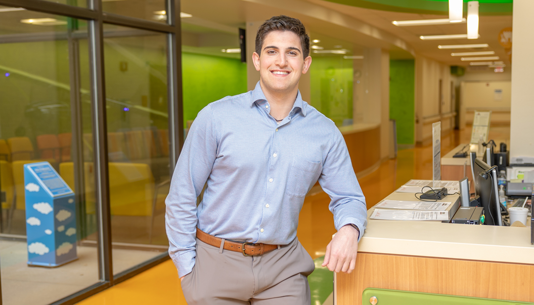
Each year, our team seeks out college students interested in pursuing a career in medicine. Clinical Orthopedic Research Assistants (CORA) are hired at Scottish Rite for Children to assist our experts in performing clinical research tasks and collaborating on research projects. CORA staff are also mentored by our experts and given valuable advice and guidance for their future. Each CORA staff member specializes in a specific area of study and works with the experts in that department, receiving a well-rounded experience in their field of interest.

What inspired you to apply for the CORA/MSRF position?
My passion and lifelong goal, since I was a kid, was to become a physician. Over the years, I have done everything I could to help achieve that goal. With my senior year at Auburn University ending, I wanted to find an experience that aligned with my goals and values, but that also helped best prepare me for applying to medical school. I am not originally from Texas, but through my college years, it became a second home thanks to some of the connections I made along the way. Whenever I expressed my goal to go into medicine, it was also followed by the same question, “Have you heard of Scottish Rite?”

When I came across the CORA position here at Scottish Rite, I knew it was the perfect match. It presented unparalleled opportunities for clinical research and shadowing and the chance to form profound career connections. Simultaneously, it aligned with my interest in orthopedics, but in a new light, where I get to make an impact on the lives of children. The most unique aspect of this position is the overwhelming support of your peers, team and Scottish Rite as a whole. They understand your goals, want you to succeed and challenge you to grow and learn as much as possible.

Have you always been interested in medicine and/or research?
Before I decided to pursue medicine, I knew that I wanted to help others. The most fulfilling thing to me was putting a smile on people’s faces knowing I impacted their lives for the better. Whether it was the numerous injuries I suffered in sports or witnessing my family battle with their own health struggles, I idolized and admired the impact physicians had on my life and the people I care about.

During my senior year at Auburn University, I worked in the Sports Biomechanics research department. I started this initially to fulfill the credit needed to graduate, but I never expected to love it as much as I did. It taught me to think critically, to question and to learn from my failures to find answers. It was this experience that sparked my passion for research and led me to Scottish Rite.

What is it like working at Scottish Rite for Children?
Working at Scottish Rite has been the most fulfilling experience of my life. I am currently a part of the Foot and Ankle team where I am surrounded by peers and mentors who challenge me each day. I not only get to learn, but I am also given the responsibility to handle tasks and problems that, although were daunting initially, have helped me grow as a researcher and aspiring physician. To top it off, I get to interact with the physicians and their patients who count on us to help give them back their childhood.

Can you share a few sentences about someone at Scottish Rite who has been a mentor to you and how they have impacted your experience? What project are you working on with that mentor?
As a part of the Foot and Ankle team, I work closely each day with Dr. Anthony Riccio. In the short time I have been here, we have worked on improving the robust Foot and Ankle and Clubfoot Registries that he developed. Additionally, I worked with him and our Foot and Ankle MSRF, Taylor Zak, to complete the Idiopathic Toe Walking Study.
Another project I am proud to discuss is one I have been able to lead in conjunction with Dr. David Podeszwa. This study will analyze the long-term outcomes of patients treated with Distal Femoral Osteotomies. In this study, I will be working closely with Scottish Rite’s Center of Excellence for Limb Lengthening and Reconstruction.

How do you think this experience will impact your career path?
In the short time I have been here, my anticipated career path has already changed. Although I have always been interested in orthopedics, since working at Scottish Rite I realized just how much I enjoy working with the pediatric population. I hope to specialize in pediatric orthopedics and continue playing an integral part of clinical research. Scottish Rite is a renowned pediatric research hospital, and I now recognize the vital role research plays in providing the best possible care to our patients.

What progress have you made towards your career goal since beginning the program?
I am currently in the middle of the medical application cycle! So far, I am grateful to have interviewed at the University of Alabama at Birmingham, the University of South Alabama, and the Uniformed Services University, and hopefully many more.

What is your favorite project that you are currently working on or have worked on at Scottish Rite?
I am working on numerous projects currently, but a few stick out from the rest. The most fulfilling study to work on is Dr. Riccio’s Clubfoot Registry and the sub studies involved with it. This study requires me to spend a great deal of time in the clinic working with Dr. Riccio and the team. While Dr. Riccio treats the kiddos, I get to monitor the pain and temperament of the babies throughout the process. In the short time I have spent here, I have developed a newfound passion for working specifically with young kids and their families.

What advice do you have for future CORA/MSRF participants?
The best advice I would give to future CORA/MSRF participants is to take full advantage of the opportunity. Scottish Rite is unique to any other hospital I have worked in. Everyone, whether they know you or not, cares. Do not be afraid to reach out for advice. Take the time to find mentors and step out of your comfort zone to develop relationships with those you admire. Especially in the CORA/MSRF programs, your peers understand your goals and want you to succeed. Use the people and resources at your disposal to pursue passions and experiences that you find interesting.

What is one thing most people don’t know about you?
In the middle of Dallas, Texas, the one thing people never expect is to find out I am an avid Washington Commanders fan!

Anything else you would like to add?
I just want to thank everybody at Scottish Rite for giving me this opportunity. It is rare to find an experience that you wake up and are excited to be a part of each day. I have learned more in these few months than ever before, and I could not be more grateful to be a part of this team!
Gracee Gets Back in the Game After The Tether™ Spinal Surgery — the First at Scottish Rite

Gracee Gets Back in the Game After The Tether™ Spinal Surgery — the First at Scottish Rite

Cover story previously published in Rite Up, 2023 – Issue 1.
by Kristi Shewmaker

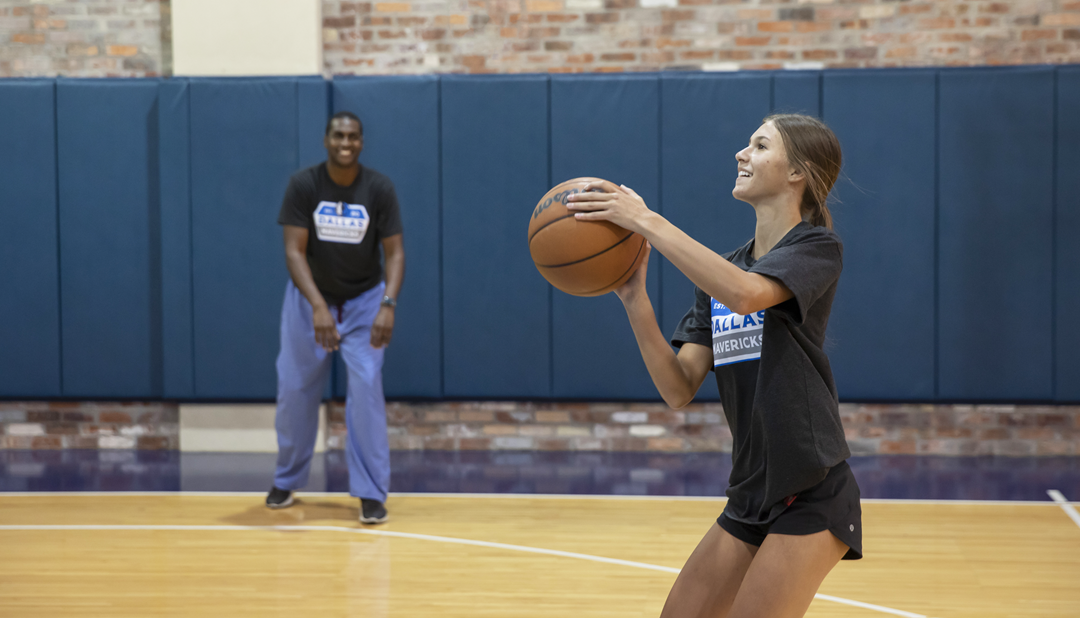
Gracee never imagined that one day she would play basketball in the Dallas Mavericks® practice gym, much less play one-on-one against her 6’5” pediatric orthopedic surgeon. As a 15-year-old high school varsity basketball player of Tuscola, she also never imagined that three months before that, she would be the first patient at Scottish Rite for Children to receive The Tether™ Vertebral Body Tethering System, a surgically implanted spinal device that is used to treat scoliosis.

Gracee grew up with basketball. “As a little girl, she went to the gym a lot because we were in the gym a lot,” says Carla, her mother. Gracee’s father, Hunter, is the high school varsity girls basketball coach. Gracee watched him coach her older sister before playing herself.

Gracee started playing competitive basketball in second grade. “We sent letters home to the girls in her class saying that we were going to meet one day a week to skill and drill,” Hunter says, “and it grew from there.” Throughout the years, Hunter coached Gracee and her team in various leagues. In middle school, Gracee joined her school team. In high school, Gracee started on the varsity team with some of the same girls that she has played with since second grade. “What I love most about basketball is the friendships that come with it,” Gracee says.

The summer before her freshman year, Gracee was seeing a chiropractor for an issue with her hip flexor, and Hunter mentioned that she had scoliosis during the appointment. A school nurse had noticed it during a routine screening when Gracee was in elementary school. Her curve was less than 10°, so her pediatrician had been monitoring it. “When she laid down on the chiropractor’s table, and I saw her back,” Hunter says, “I was like, ‘Oh my gosh!’ We didn’t realize to what degree the curve had gotten.”

Hunter contacted Scottish Rite for Children for help, and there, Gracee was diagnosed with adolescent idiopathic scoliosis. This condition occurs in patients ages 10 to 18, and the cause is unknown. Her curve measured 42°. “I was shocked,” Gracee says. “I didn’t think it would be that bad.”

Initially, Gracee wore a brace to prevent further progression and to try to avoid surgery. However, within months, her curve progressed to more than 50°, which experts call the surgical threshold — the point when surgery is recommended to stop a curve from getting larger. “In a certain subset of kids wearing their braces, the curve still progresses,” says Jaysson T. Brooks, M.D., pediatric orthopedic surgeon. “They have so much growth potential left, their spine overpowers the brace.”

When Gracee and her family first met Dr. Brooks, they began discussing treatment options in anticipation of the need for surgery. Dr. Brooks presented two surgical options, including spinal fusion, which he calls “the gold standard treatment,” and The Tether™, a new non-fusion surgical device he had used previously to treat scoliosis but had not been used at Scottish Rite.

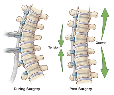
In a spinal fusion, screws are inserted into channels of bone surrounding the spinal canal. Two rods are secured into the back of the screws with set caps and tightened with a torque wrench. Bone graft is placed along the implant to help the vertebrae fuse together. This procedure stops the growth in the previously curved part of the spine and prevents the curve from coming back.

When using a tether, a flexible cord is attached with screws to the vertebrae on the side of the spine that curves outward, which is also known as the convexity of the curve. The tether slows the growth on the outward side of the curve while allowing the inward side of the spine to continue growing. During surgery, the flexible cord is tensioned, which corrects the curve to some degree. As a child grows, the spine grows straighter because the tether guides it into the correct alignment. The Tether™ is a non-fusion implant that allows children to maintain the majority of their natural spine flexibility
 
“Gracee was a good candidate for this procedure because her curve is flexible, and she was still really young from a bone standpoint, or skeletal maturity,” Dr. Brooks says. “The younger your skeleton is, the higher chance you have of getting taller, and using a tether requires you to be able to grow taller because it capitalizes on that growth.”
 
Over several appointments, Dr. Brooks discussed the pros and cons of each surgery, the difference in incisions, how X-rays look when rods versus a tether are attached to the spine, and the potential downsides and risks. “He did a really good job explaining the difference between the two,” Gracee says. “He also did a really good job of making me feel calmer.”
 
“There wasn’t a lot of anxiety on our part about which way to go,” Hunter says. “In my mind, vertebrae are meant to be flexible. If you can keep it the way God designed it, I think you’re better off. Obviously, there are times when the fusion is the only answer, but with this being another option, we wanted to try it.”
 
Gracee’s family chose this device because they wanted to maintain the overall flexibility of her spine as well as endure a shorter recovery period, so that Gracee could get back to playing basketball as quickly as possible.
 
“They are very intelligent and very pragmatic to approach a procedure that is this new and say, ‘I think this is going to be the right thing for my daughter,’” Dr. Brooks says. “They were mature enough as a family to say, ‘we’re okay with the potential consequences, but we think that the benefits outweigh the risks.’ You couldn’t have asked for a better family to be the first family for The Tether™ at Scottish Rite.”
In April of 2022, Gracee underwent surgery. “I wasn’t that nervous going into it,” Gracee says. “When I got out, I was excited to finally be good. I was excited about it.”
 
“The whole time, I felt like she was in very good hands,” Carla says. “Dr. Brooks’ knowledge is one thing, but he is very compassionate and caring as well. I truly felt like he loved my child.”

Two weeks later, Gracee went back to school. After six weeks, she played her first post-surgery game, and after three months, she found herself at the Dallas Mavericks® practice gym playing basketball with Dr. Brooks.

“It was a really cool experience,” Gracee says. “Dr. Brooks did pretty good.”

The feeling was mutual for Dr. Brooks. “It was a special opportunity for sure,” Dr. Brooks says. “I was pretty good at basketball in high school, but I’m super out of shape now. The only thing I had to my advantage was my height. Where she beat me in speed, I used my height.”
 
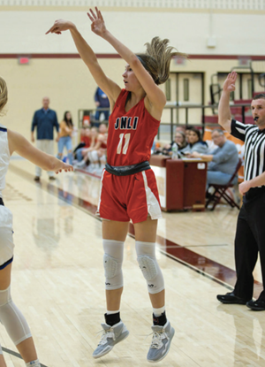
Gracee has played shooting guard for as long as she can remember. Hunter explains that she shoots in the 40% range from the three-point line. “Gracee’s a good shooter because she’s up at the gym at 5:00 or 5:30, three or four mornings a week working on her shots,” Hunter says. “Our basketball program is highly competitive. They’re ranked No. 2 in the state.”
 
In December, Gracee’s team won the Whataburger® Basketball Tournament, an invitation-only tournament in Fort Worth. When it was over, five players out of approximately 160 girls were chosen for the alltournament team. Gracee was one of the five.
 
“Going from being on a surgery table in April to being able to do that in December is just phenomenal,” Hunter says. “Gracee’s always been a spunky kid. There wasn’t much spunk a year ago, but we’re seeing it come back.”

Today, Hunter says that they have no regrets about choosing tethering, but he knows that it is still early. During surgery, Gracee’s spine was corrected to a little more than 30°. The goal is that the tether will continue to straighten her spine as she grows.

“If Gracee had walked into my clinic with a 30° curve, she would not have gotten surgery because 30° isn’t big enough,” Dr. Brooks says. “In the end, if nothing changes, it’s still a win because she has a curve that is stable. There are lots of kids walking around with 30° curves who are living full, successful lives.”

Hunter says that they would be happy if her curve stayed where it is and ecstatic if it improves even more. “We’re just fortunate that we’re talking about a scoliosis curve that is manageable,” Hunter says. “When you see what other families at Scottish Rite are dealing with and the care that you see being administered to them, it’s as close to heaven on earth as you think you’ll ever see.”
 
“Everyone works so hard to make the kids at Scottish Rite feel at home, to make me feel at home,” Gracee says. “I wasn’t scared to be the first kid at Scottish Rite to get The Tether™. I like being groundbreaking. I like being the first.”
 
“And, our experience at Scottish Rite,” Hunter says, “has been nothing but first-class.”

Read the full issue.

Infantile Clubfoot Deformity

Infantile Clubfoot Deformity

Clubfoot deformity occurs in one in 1,000 live births, making it the second most common congenital deformity of the foot (after postaxial polydactyly) and one of the most encountered infantile musculoskeletal congenital differences. In contrast to other common infantile foot deformities, such as metatarsus adductus and calcaneovalgus deformities, a clubfoot is not a normal foot that has been “packaged” into an abnormal position. Rather, a clubfoot is a true congenital deformity. There are structural differences inside the foot that create the outward deformity. The shape of the tarsal bones is not the same as that of a child with a normal foot. The elasticity of the soft tissues is much less than in a child with a normal foot, and arterial vessels are often absent or aberrant. Some of the extensor muscles in the leg have been shown to have an exponentially higher number of contractile elements (actin and myosin filaments) and are thus able to generate force at what is essentially a superhuman level.

Despite our understanding of the underlying anatomic and histologic differences in clubfeet, the exact etiology of the deformity remains unknown. Although genetic aberrancies have been identified that are associated with the clubfoot, these appear to be a result of somatic mutations, and thus, the deformity does not appear to run in families with any identifiable pattern of inheritance.

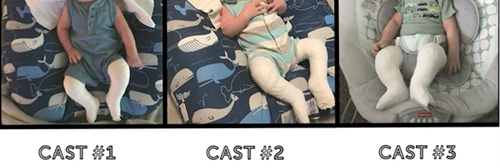
In approaching the treatment of a clubfoot, it is essential for families and providers to understand the presence of these congenital differences because our method of treating clubfoot cannot alter any of them. We can’t change the shape of the bones. We can’t improve the elasticity of the soft tissues. We can’t change the way the blood vessels move into the foot, and we can’t change the power with which certain muscles fire. Therefore, the goal of clubfoot treatment isn’t to create a normal foot. Rather, the goal of treatment is to take a structurally abnormal foot, which is also in an abnormal position, and through a series of casts applied weekly, slowly convert into an abnormal foot structurally that is ultimately in a near normal or completely normal position. Also, this method limits the need for surgery, which creates scarring and stiffness, thereby preserving as much motion of the foot as possible.

The Ponseti method of treatment is the gold standard of care worldwide for clubfoot deformity. This involves weekly serial casting of the involved foot or feet, with each cast applied in a manner to correct a different component of the clubfoot deformity. These casts are very effective at correcting every component of the clubfoot deformity with great success, save for the contracture of the Achilles tendon, which drives an oftentimes rigid equinus (plantarflexion) deformity of the ankle. Approximately 90% of clubfeet require a percutaneous transection of the Achilles tendon. This is performed in the clinic with topical anesthesia in children aged less than 3 months to overcome this component deformity. Following the heel cord tenotomy, the ankle can be acutely dorsiflexed to at least 10 to 15 degrees, and that position is maintained in a final cast that is kept in place for three weeks.

On average, it takes about four weeks of weekly casting to correct the component deformities of the cavus, hindfoot varus, and metatarsus adductus. That brings the foot from what is essentially an upside down and turned inward position to a right-side-up and turned-out position. At that point, if the ankle remains in equinus, this deformity is corrected with a heel cord tenotomy and a final three-week cast, resulting in a total time to achieve full correction of about seven weeks. As we ideally begin treatment within one or two weeks of life, most deformities are corrected by or before 2 months of age.

It cannot be overstated that deformity correction is just the first part of the treatment of a clubfoot deformity. Maintaining that correction is essential and, in many regards, more difficult. Seven weeks in a cast simply cannot overcome months of intrauterine gestation and the various congenital differences present within these feet. Without a means of maintaining correction, corrected clubfeet will universally and quickly revert to their initial position of deformity.
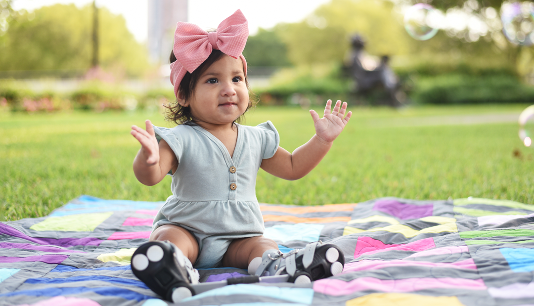
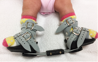
Maintenance of correction is achieved by keeping the ankles dorsiflexed and the feet externally rotated in what is typically called a “boot and bar” construct. While there are a variety of Ponseti-type braces available, most consist of soft silicone lined shoes with soft suede straps. Regardless of whether the deformity is unilateral or bilateral, these shoes are applied to both feet and connected to each other by a bar. The bar holds the shoes at 60 to 70 degrees of external rotation, and the shoe straps hold the ankles in neutral dorsiflexion.

Though bracing protocols vary to some degree, our protocol mirrors that agreed upon at the International Clubfoot Congress and consists of brace use for 23 hours a day until the child begins to pull to stand and 12 hours a night thereafter until age 4. The braces do not interfere with the achievement of developmental milestones, such as sitting up, rolling over, crawling or standing. The transition from full-time to part-time use is based entirely on the fact that once a child is able to pull to a standing position, they will soon spend more time on their feet. At that time, their own body weight will help to keep their feet flat on the ground. Until that time, babies spend most of their time lying down and sitting, during which the feet are held in a relaxed position. That can result in tightening of the heels cords and a recurrence of equinus.

Bracing is most difficult on parents for the first two weeks after the child comes out of their post-tenotomy cast. After seven weeks of casting, having anything other than cast padding surrounding their feet feels different and can cause fussiness. Parents are counseled extensively about this so they can anticipate and develop strategies to keep the braces in place despite frustration on the part of the child.

As mentioned, the use of the brace and the prescribed protocol is essential to long-term treatment success. If the braces aren’t used as recommended, the risk of recurrence of the deformity approaches 100%. These recurrences can be much more difficult to manage in older children than during infancy. It typically requires repeat casting and general anesthesia for either a repeat heel cord tenotomy or a more invasive surgical intervention.

Unfortunately, even when perfect correction is achieved with casting, and parents are compliant with brace use, a percentage of these deformities still recur and require additional treatment due to the structural congenital differences present. Research has demonstrated that with parental bracing compliance, about 80% of clubfoot patients never need any additional treatment. However, despite everyone’s best efforts, 20% of children still need some additional treatment due to recurrence. These recurrent deformities are most commonly the result of an imbalanced muscle pull across the foot due to a relatively overpowered tibialis anterior tendon. When relatively overpowered, this muscle, due to its insertion along the medial aspect of the foot, supinates the foot during dorsiflexion. That results in the touchdown of the lateral border of the foot during the initial stance phase of gait. This supination, in turn, slowly drives the foot inward and into further supination with weightbearing. Such recurrences are easily managed with a brief period of repeat casting to correct any relapsed deformities. That’s then followed by a transfer of the offending tendon to the dorsum of the foot to remove that supination moment during dorsiflexion.

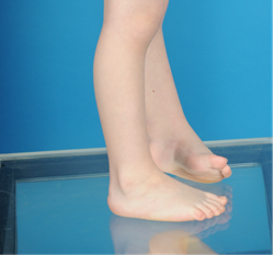
With an experienced team of providers and parental dedication to brace use, these initially dramatic foot deformities can be corrected beautifully and without any long-term deficits or disabilities. However, it is important to educate parents early regarding certain differences in the foot or feet following treatment. A clubfoot is always smaller than a normal foot, sometimes half to a full shoe size smaller than an unaffected contralateral foot. In addition, the calf musculature of children with clubfoot tends to be smaller, though this size difference does not appear to affect strength. These differences are strictly cosmetic and do not alter function. The goal of clubfoot treatment is to allow a foot or feet to sit flat to the ground, have as much flexibility as possible, allow the child to wear any shoes they want, play any sports they want, have any job they want, and do whatever they want with the foot. In achieving this, their corrected clubfoot will function no differently than a normal foot and will never limit a child in any way in any activity they desire.

Download the PDF.

Dr. Anthony I. Riccio is a pediatric orthopedic surgeon and the Director of the Center for Excellence in Foot at Scottish Rite for Children.

Waterproof Cast 101

Waterproof Cast 101

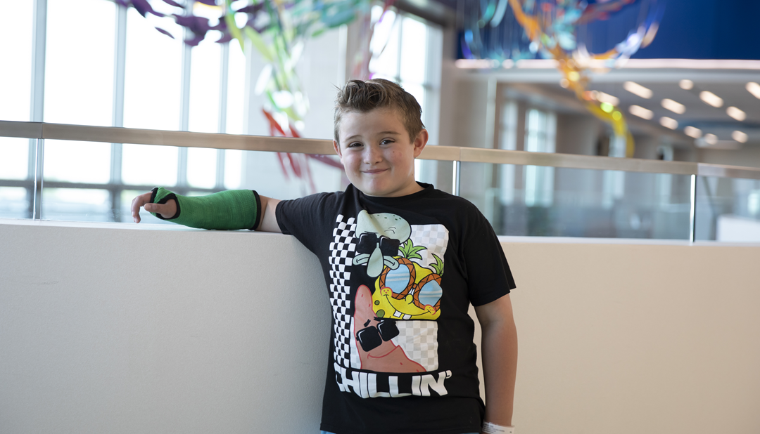
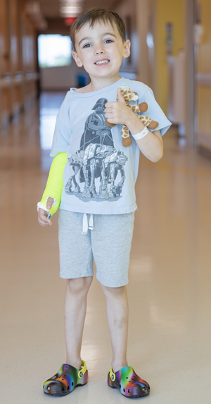
Casts are made of two layers — a soft inner layer and a hard outer layer. The inner layer of a waterproof cast uses a special type of lining that allows it to get wet when bathing, showering or swimming.

Does a waterproof cast have any risks?
Every time a waterproof cast gets wet, it needs to completely dry. If the skin inside the cast stays wet for a long period of time, it can become irritated. If your child develops a rash or burning sensation while in the cast, notify your clinic immediately.

Waterproof cast care

  • A waterproof cast needs to get wet daily.
  • After your cast gets wet, make sure all of the water exits the cast by moving your arm/leg around.
    • Your natural body heat will evaporate the rest of the water in the cast.
    • You may also use a hair dryer, on the cool setting, to finish the drying process in areas like the elbow crease or heel of the foot.
  • Following bathing and swimming, flush the inside and outside of the cast with a forceful stream of clean water only.
  • If the cast gets itchy, dirty or smelly, you may rinse mild soap through the cast and then thoroughly flush it with clean water.

What else should I know about caring for a child in a waterproof cast?

  • Follow directions and activity restrictions given by your provider.
  • See cast care patient education for more information.
  • You may swim in lakes and oceans with the waterproof cast, but it can be difficult to remove sand, dirt and other small particles from inside the cast. If particles are left in the cast, they can cause skin irritation and discomfort. Please be sure to flush the cast thoroughly with clean water until all particles are removed before allowing the cast to dry.

Learn more about our Fracture Clinic.

The Growing Athlete’s Hip: How to Prevent Problems Today and Tomorrow

The Growing Athlete’s Hip: How to Prevent Problems Today and Tomorrow

Download a PDF of this summary.

In this program, our pediatric orthopedic and sports medicine experts described how the skeletal development of the hip is affected by repetitive and extreme movements inherent to athletic activity. The changes, in some cases, can be permanent. Keep reading to learn what we know about preventing irreversible changes and treating symptoms of these sport-related hip conditions.

Apophysitis and Apophyseal Fractures in the Hip and Pelvis

Apophysis is a normal bony outgrowth that arises from secondary ossification centers. The bone fragment will ultimately fuse with the primary bone. The apophysis contributes more to the shape of a bone than the longitudinal growth. Until the ossification center fuses, also referred to as the point at which the “growth plate closes,” the tendon or ligament attached to the apophysis can pull and cause pain in the soft cartilage in the apophysis.

Hip and pelvic apophyses that are vulnerable to acute or overuse injuries are located at the ischial tuberosity, the iliac crest, the anterior superior iliac spine (ASIS) and anterior inferior iliac spine (AIIS). An apophyseal avulsion fracture. An anterior-posterior view of the pelvis is helpful when evaluating complaints in the pelvis so contralateral comparison can be made.
Risk factors for injury includes:

  • Tight muscles and muscle groups
  • Early in the sports season
    • Change in activity from sedentary to active
    • Sudden increase in intensity or duration of training or competition
  • Ignoring activity-related pain
  • Minimal recovery from workouts
    • Year-round training
    • Lack of cross-training
    • Overtraining

Treatment for these conditions is most often nonoperative and is centered around protecting the area involved. Rest, protected weight-bearing, gentle passive ROM and gradual return to play are necessary elements of the plan. Healing and symptom resolution may take 12 weeks or more and radiographic healing is not required prior to returning to sports.

Internal and External Snapping Hip

Athletes may report “popping” in the hip.

If you can see it, it’s likely coxa sultans externus, external snapping hip. This is a condition of the iliotibial band popping over the greater trochanter on the lateral side of the femur. Runners may complain of this when running or walking, and they may describe that it “pops in and out.”

If you can hear it, it’s likely coxa sultans internus, internal snapping hip. This occurs when the iliopsoas muscle, deep in the groin, causes painful popping. This condition is often seen in dancers and tumblers. Treatment includes hip flexor stretching and activity modification.

Femoroacetabular Impingement (FAI)

An overuse injury seen in adolescent and young adult athletes in the hip can be caused by changes in the shape of the femoral head-neck junction (Cam-type) or the acetabulum (Pincer-type). These changes can cause pinching and tearing of the labrum, the soft tissue surrounding the acetabulum that acts to deepen the socket. Early injury from impingement can cause premature hip arthritis. Therefore, this condition is continuing to get more attention with the goal to prevent deformity and consequences.

How does a Cam-type deformity develop?
The femoral head collides prematurely with the acetabulum. The impact causes a change in the shape of the head from being spherical to being more “cam” shaped, or oblong. These may develop secondary to another medical condition in the developing hip, such as:

  1. Slipped capital femoral epiphysis (SCFE) is seen in approximately one in 10,000 may occur and result in avascular necrosis of the femoral head.
  2. Perthes disease – rare condition affecting blood flow in the hip and causes deformity.
  3. Trauma or fracture

In athletes, there is not a primary condition like those listed above. Therefore, idiopathic Cam deformities have been identified in teenage athletes who participate in soccer and other sports. Younger players studied do not show this condition, so the window of opportunity and the exacerbating activity are being studied more closely. Shearing forces may be occurring at the physis to protect the bone, but ultimately may be causing changes in the growth plate and therefore the shape of the femoral head.

Can this be prevented?

Early conversations are looking at the parallel occurrence in the shoulder and elbow in baseball players. Evaluation of the dosage of activity, such as pitch counts in baseball, have been implemented to preserve the anatomy and improve performance in elite athletes. For now, working on proper mechanics and activity modification in adolescence may be our best tools to prevent this deformity.

Considerations and Components of a Hip Injury Prevention Program

Factors that must be considered to prevent hip injuries in adolescent athletes include:

  • Open growth plates
  • Peak height velocity (PHV)
  • High volume of training particularly with loading in rotational and axial movements
  • Sport-specific end range of motion demands
  • Explosive and eccentric demands

Modifiable factors may include:

  • Muscle imbalances
  • Muscle weakness
  • Inflexibility
  • Poor technique
  • Sport-acquired deficiencies
  • Joint instability
  • Overtraining

Five Domains of Injury Prevention Strategies of the Hip

  1. Training Load Management
    Higher incidence of athletic hip pain found with athletes who specialize in a single sport before high school and participate in regular training at earlier ages and four times per week before the age of 12. Recommendations include sampling a variety of sports rather than specializing, monitoring workload, neuromuscular training programs and taking rest breaks from sport (two to three nonconsecutive months/year).
  2. Hip Mobility During Rapid Growth
    Through stretching, dynamic warm-up and eccentric training, hip tissues can stay flexible. Progression of eccentric training can improve the length-tension curve to improve performance and resist injuries.
  3. Motor Control and Stability
    Hypermobility and poor motor control need to be addressed with strategies that improve core stability and teach foundational movement patterns for sport-related movements, such as jumping and landing.
  4. Strength to Improve Imbalances & Specificity
    Once mobility and control are addressed, strengthening can occur. Eccentric adductor & abductor strength can be improved by combining activities, such as the Copenhagen plank and a Nordic Hamstring exercise. Looking for sport-specific strengthening tasks.
  5. Sport-Specific Movement Mechanics
    The culmination of these strategies is executing the sport-specific movement patterns with all of the fundamental movement competence and technical accuracy to ensure safety. Whether the sport demands jumping and landing on a court, changing direction at high speeds on the ice or holding extreme postures on a balance beam, the steps follow a standard pattern.

Implementing Hip Injury Prevention Programs

With confidence that many of these elements are modifiable due to neural plasticity of youth athletes before and during growth, making an effort to prevent injuries is appropriate. Research will continue to define the right and wrong approaches; however, we have some tips that are generally accepted. To avoid detraining, it is recommended to perform activities two to three times per week, approximately 20 min duration, up to 60 min for at least six weeks. It is important to implement it prior to the beginning of a season. Qualified instructors and supervision for continued implementation of the proper techniques are crucial elements of a safe and successful program.

Learn more about hip health in dancers.

This is a summary of a presentation in a monthly series for medical professionals called Coffee, Kids and Sports Medicine. Through events like these, Scottish Rite for Children experts share their experience and knowledge with others to ensure young and growing athletes are getting the best care in every environment.