Share Your Story: Cambryn Mannos – Redefining Myself

Share Your Story: Cambryn Mannos – Redefining Myself

Blog written by Cambryn, and Cambryn’s mom, Amy.

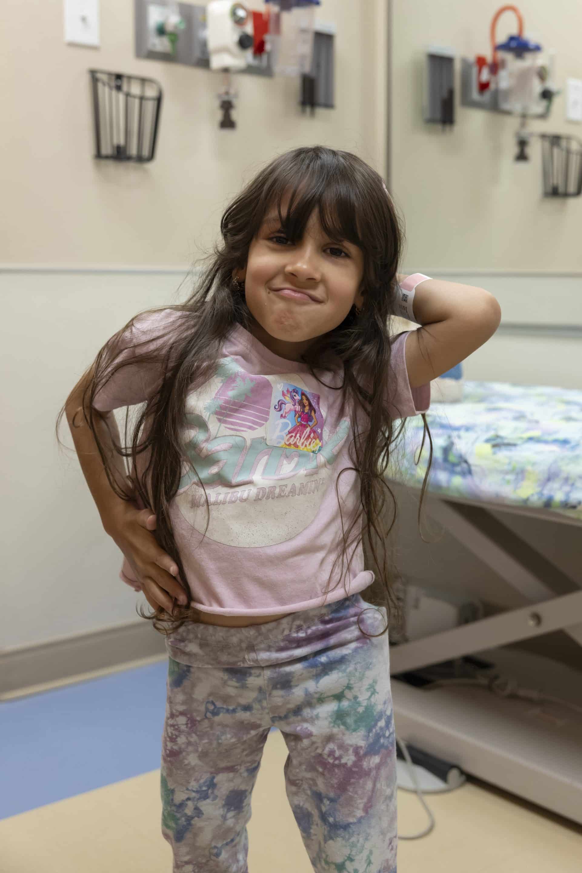
Go back a year and you could find me, Cambryn, age 9, training with the best team and best coaches in all of New Mexico. My sport is gymnastics — the sport of all sports that defines toughness, perseverance and dedication. Winning competition after competition, I was defined as the best in my age group, hardworking, fearless, fireball, hotshot and destined to be counted among some great gymnasts. I was born into gymnastics, raised at Gold Cup Gymnastics School and the true definition of a “gym rat” with two older siblings competing. The gym is where you could find me all day, every day! Until the day it all came crashing down. It began with hip pain surging with every landing and movement in any backwards motion, including back walkovers, back tucks, back handsprings and back layouts.

My ability and stability were gone, just like that. I visited my orthopedic doctor right away at the request of my head coach, and on the X-ray, the doctor saw a fracture on the head of my left femur. An MRI was ordered, and while waiting, my ability to walk was gone by Christmas Eve. My MRI was now on stat order, and BAM, with all its glory, my MRI lit up black spaces through the head of the femur, showing avascular necrosis and Legg-Calve-Perthes disease, big words that I did not understand.

I struggled to find words to define my thoughts. I was referred to another orthopedic doctor in pediatrics, and “wait and see” was the advice. Then, my hip collapsed. All that I was and all that I thought I could be vanished, my definition of myself was gone. I lost it all! And yet again, “wait and see” was the approach. Now I needed a wheelchair, except after three months, I was still on a waitlist to get a wheelchair. 

Nothing seemed to happen but waiting, and I could see nothing in my future to be but that same blackness or death of what I was and what I could be. Gymnasts don’t “wait and see,” they are proactive and always in a constant state of motion. This is where my redefining journey begins with Scottish Rite for Children. With help from my teammate’s dad, who is a doctor, we found Dr. Harry Kim and his nurse Ms. Kristen. Their knowledge and dedication are a priceless need for us Perthes kids. They gave light to the blackness I saw because they had a game plan! 

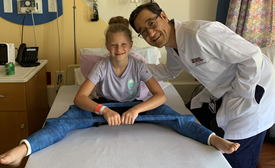
Now, the beginning of the fight to redefine myself is not my proudest moments. I have had to do some soul-searching and letting go of a lot of pride and previous definitions of myself to build the “Perthes” me. Because at first, I freaked out at Dr. Kim. “He wants what? Six weeks in a huge cast? Nope, not doing that,” was my response. Yet, this summer, I did the tenotomy and petri casting, and I survived and even thrived with some new adventures. Dr. Kim is amazing, kind, patient and willing to fight my battle with me. Because of him, I know that Scottish Rite for Children is the place for me to get better and be a part of something bigger. You become family at Scottish Rite. I gained family. My care each time I go has been top of the line care. It’s worth the 1,400-mile trip to see my Scottish Rite family and to belong to something big, progressive and healing. 

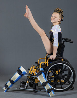
Today, I find myself in stage 3 of Perthes disease, rebuilding new bone and redefining myself in all that I lost and in all that I have gained. I can grieve what I lost, and I can work hard to regain most of what was lost, but mostly I can seek moments that empower myself so I can empower others. I recently took photos for gymnastics. At first, I was embarrassed at the thought of gymnastics photos with a wheelchair. But the photographer, Kevin Lange, helped me see a bigger picture. I embrace Perthes disease because it is a part of me now, and I can’t change that, but I can choose what I do with it. I choose to find joy and strength in my Perthes journey. To me, Scottish Rite means the chance to regain and redefine myself! It means a new family of people who care about my health. It means everything to my future self who cannot fight this battle alone.

Thank you, Dr. Kim and Ms. Kristen, for your time and energy, for your dedication and expertise. 

Other than gymnastics, I enjoy rock climbing, math, animals, bike riding, hiking and art.

Photo Credits to Kevin Lange


DO YOU HAVE A STORY? WE WANT TO HEAR IT! SHARE YOUR STORY WITH US.
Brock’s Brigade – Fighting Perthes Step by Step

Brock’s Brigade – Fighting Perthes Step by Step

Cover story previously published in Rite Up, 2022 – Issue 3.

by Kristi Shewmaker
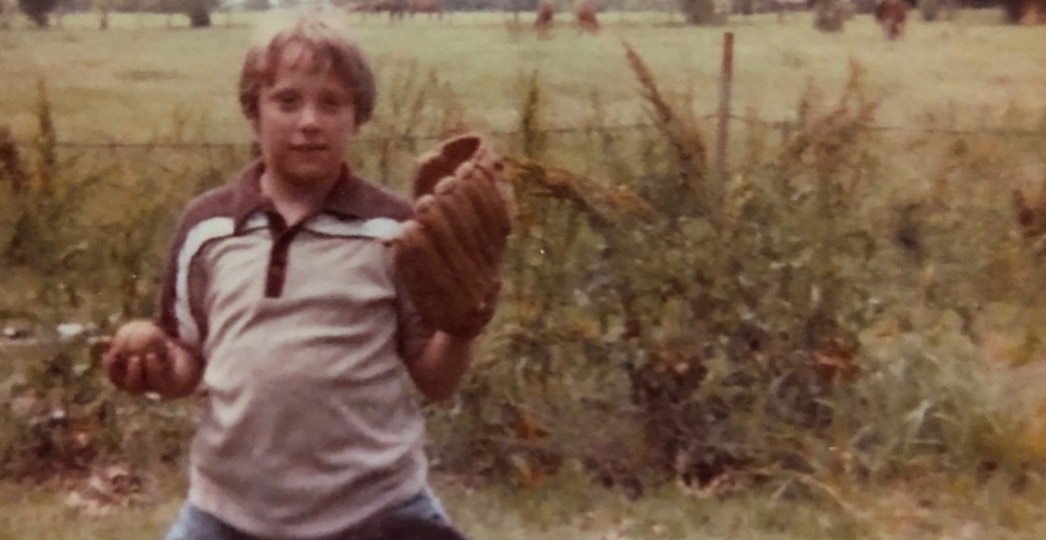
Kickball, wiffle ball, four square, taking hikes and riding bikes are a few of the activities 10-year old Brock, of Lee’s Summit, Missouri, enjoys. But, his first love is baseball.
 
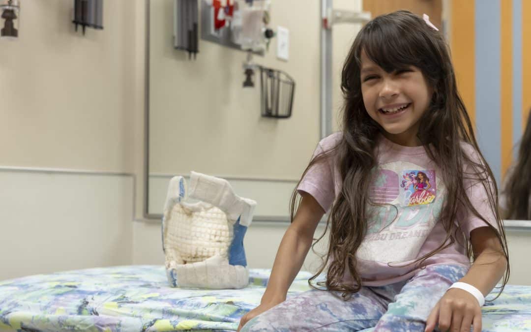
“Baseball is his world,” his mother, Rachel, says. “He has played competitive baseball since he was 4.” In the spring of 2021, Brock played shortstop for a local team called the Baseknocks until midseason when he was diagnosed with Legg-Calvé-Perthes disease (Perthes), a rare childhood hip disorder, that temporarily took him out of the game and into a wheelchair.
 
Brock’s symptoms began with a pain in his groin. Then, he started to limp. “It was really bad,” Rachel says. “He couldn’t not limp.” She took Brock to visit his pediatrician who examined him but found nothing obvious like a broken bone. The doctor suggested trying physical therapy. “We thought maybe he had pulled a muscle,” Rachel says. After almost two months of physical therapy, Brock was still limping, and Rachel noticed that the thigh muscle in his right leg was two inches smaller than the thigh muscle in his left leg. “That was pretty alarming to me,” Rachel says. “I thought, ‘There’s something going on. It’s not just a pulled muscle.’” Later, an X-ray revealed that Brock had Perthes, a disease unknown to the family.
 
STEP 1: FINDING AN EXPERT

Perthes disease is a hip disorder that primarily affects the ball of the hip joint. The ball, or femoral head, is the upper part of the thighbone, or femur. The femoral head is normally round and fits inside the round socket of the pelvis. Perthes disease occurs when part or all of the femoral head loses blood supply. Without adequate blood flow, the femoral head bone dies. Over time, the body removes the dead bone and replaces it, initially, with softer bone. This bone is weaker, and the femoral head is more likely to collapse into a flattened position leading to deformity of the hip.
 
Perthes is rare, affecting approximately 15 children per 100,000 and is more common in boys than in girls, with a ratio of 5:1. The cause of Perthes is unknown, and currently, there is no cure. It typically occurs in children between 4 to 8 years old. Brock was diagnosed just before he turned 9.
 
“When you’re diagnosed with something that’s rare, and there’s not a ton of information, you feel like it’s a death sentence,” Rachel says. “I remember crying for the first three or four days because there were no answers, and no doctor could get us in. It was like a big, giant question mark.”
Rachel asked a friend, a physical therapist in nearby Kansas City, if she could recommend someone. Her friend said, “If it were my child, I would go see Dr. Kim at Scottish Rite.”
 
“Late at night, I sent a message to Scottish Rite and received a call the next day,” Rachel says. “Dr. Kim had an opening the day that we were going to be driving back through Dallas after a spring break trip to Galveston. It was clear that it was meant to be.”
 
STEP 2: GETTING EXEMPLARY CARE

A leading expert in Perthes disease, Harry Kim, M.D., M.S., is a pediatric orthopedic surgeon and director of the Center for Excellence in Hip at Scottish Rite for Children. Dr. Kim met with Brock and his family and confirmed the diagnosis.
 
“A perfusion MRI shows how much of the femoral head has blood flow,” Dr. Kim says. “Normally, it should be 100%. In Brock’s case, about 90% had no blood flow, which caused about 90% of his bone to die. His case was severe based on the amount of bone death and his age.”
 
Children ages 6 and younger tend to have better outcomes from Perthes disease because they have greater potential for developing new bone. Also, if half or more of the femoral head dies, the potential for regrowth without deformity is lower.
 
During the family’s initial visits, Dr. Kim and his team, including registered nurse Kristen Odom, explained the disease and Brock’s specific case and outlined the treatment options.
 
“When we came out of that first appointment, Brock said the best thing was that Dr. Kim looked at him, talked to him and asked him questions about how he was feeling, rather than just talking to me,” Rachel says. “It’s so important when you’re scared and uncertain, especially when you’re a kid, and the doctor makes you feel like you truly matter.”
 
After returning to Missouri, Rachel had more questions as the family processed the information. “Kristen spent an hour and 45 minutes on the phone with me,” Rachel says. “When I hung up, I told my husband we won’t ever go anywhere else. That kind of care simply doesn’t exist. When you’re dealing with a rare disease and your baby, that care is priceless.”
 
In May of 2021, Dr. Kim performed surgery on Brock’s right hip. “Brock presented with a mild collapse or deformity of the femoral head,” Dr. Kim says. “Without treatment, it would have degraded much further. We intervened before further collapse occurred.”
 
During the procedure, Dr. Kim cut the bone and positioned it so that he could tuck the femoral head securely into the hip socket. This containment procedure allowed the blood flow to come back naturally. He stabilized the bone with a metal implant that was removed in a later surgery. Over time, the body would remove the dead bone and generate new bone. To ensure healing, Dr. Kim prescribed a controlled weightbearing regimen, meaning Brock was unable to put any weight on his leg.
 
“He couldn’t play baseball, couldn’t run up the street to a friend’s house, couldn’t ride his bike,” Rachel says. “He went to school in a wheelchair, and kids stared at him and asked questions. We thought about ADA (Americans with Disabilities Act) accommodations everywhere we went. We had to rethink everything.”
 
STEP 3: FIGHTING FOR OTHERS

Despite all of these changes, Brock has taken it in stride. “He is my hero,” Rachel says. “Has he complained? Absolutely. Did he hate certain minutes, days and weeks? 100%. But overall, he was relieved to have an answer and a plan and no more pain. He has handled it better than I could have ever imagined.”
 
Last Christmas, Brock wanted to express his gratitude by designing and selling T-shirts as a fundraiser for Scottish Rite. “We called ourselves ‘Brock’s Brigade,’” Rachel says, “and put ‘His Fight Is My Fight’ on the front.” When the family traveled to Dallas for Brock’s second surgery in December, they presented a check for $1,000 to his care team. “Dr. Kim has really made an impression on Brock,” Rachel says. “This was Brock’s way of giving a little back so that hopefully it’s easier for kids in the future.”
 
“This family is so special,” Dr. Kim says. “They have gone through such difficulty, yet they want to help others improve their knowledge and support research. They are not just thinking about themselves but thinking about others.”
 
For more than a year, Brock completed a series of progressive exercises to restore muscle strength and range of motion. He went from using a wheelchair to bearing more and more weight on his crutches. In August, the answer the family had been holding their breath for finally came. Brock was given the all clear to walk.
 
“After we got the A-OK, Brock took his first walk with his dad and his brother,” Rachel says. “They do these “football walks” where his dad throws the football, and they go up ahead and catch it. They got to do that for the first time in a long time.”
 
Brock is especially excited for the day when he is cleared to play the sport he loves most. “Dr. Kim felt confident that Brock would be able to start winter practices and be 100% for spring baseball, which has been his goal from day one,” Rachel says. For now, Brock is easing back into his active life. He walks his dog, Pepper, and plays on the playground with his friends.
 
“The Scottish Rite team has been the biggest blessing to our family,” Rachel says. “Even being eight hours away and having to drive and fly multiple times throughout the last 18 months, I would drive four days to get to Scottish Rite. Our experience has been nothing short of amazing.”

Diagnosing, Referring and Treating Newborns with DDH

Diagnosing, Referring and Treating Newborns with DDH

Watch the lecture on YouTube or read this summary to catch the highlights.

Download the PDF.

This is a summary of a presentation for medical professionals that focuses on developmental dysplasia of the hip, or DDH. Presented by William Z. Morris, M.D., the seminar dives into everything medical professionals need to know about evaluating and treating DDH in newborns, helping physicians recognize the condition and respond earlier.

DDH is a common condition that occurs in about one in 100 infants. The condition is characterized by a shallow acetabulum and/or under-covered femoral head in the hip. It can occur due to a malformation of anatomic structures that have developed normally during the embryonic period and ranges in severity from physiologic immaturity to subluxation to frank dislocation. The presentation covers the epidemiology of DDH and its risk factors.

Dr. Morris provides updated guidelines for selective ultrasound screening for high-risk infants and includes data from his recent publications and presentations at national conferences. The presentation covered a full DDH screening and physical exam, showing providers exactly how to look for signs of DDH in newborns. He explains that physical findings fall on a spectrum and vary with the severity of the pathology and the age of the child. The presentation includes a detailed video of a newborn physical exam, showing participants hip-specific tests that can be performed to identify even subtle signs of dysplasia.

Email medicalprofessionals@tsrh.org to request access to the full exam video.

Imaging is a valuable tool in helping to diagnose DDH, but Dr. Morris shares why it is best to wait until the patient is 6 to 8 weeks of age in cases of screening ultrasounds for stable hips,  using facts and figures to illustrate this reasoning. He recommends ultrasounds at 6 to 8 weeks of age, which reduces false positive rate, and X-rays after 6 months of age once the hip has undergone sufficient ossification.

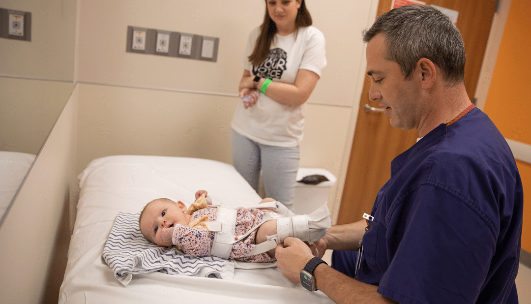
The presentation continues with Dr. Morris describing treatment protocols for DDH. For many, primary treatment for DDH begins with a Pavlik harness for six to eight weeks. He shares what to watch for with this treatment and its success rate using granular data in order to arm primary care physicians with data that can be used to reassure families once the diagnosis is made. He then talks about further treatments, including hip abduction brace, closed or open reductions and spica cast, and in which cases each may be used.

Finally, Dr. Morris shares vital information about DDH prevention, such as healthy hip swaddling, the use of proper sleep sacks and the correct use of baby carriers and how each of these can contribute to DDH in newborns.
Dr. Morris encourages physicians to refer patients early and often in cases of suspected DDH, know the risk factors and help parents with prevention techniques. He stresses that in most cases, nonoperative treatment is very successful, especially when the condition is caught early. Pediatric physicians and their patients can greatly benefit from Dr. Morris’ expertise with DDH, learning everything physicians need to know to provide their smallest patients with the best care.

Preventing Hip Problems for Your Baby

Preventing Hip Problems for Your Baby

Newborns need a lot of care, and that means plenty of visits to the pediatrician during the early months. One thing your pediatrician will carefully screen for is developmental dysplasia of the hip (DDH), a common condition that young babies are especially susceptible to. Learn more about DDH, its risk factors, tips on how to prevent the condition and guidelines on how to spot hip-safe baby accessories from our experts.

Could my baby have DDH?
DDH occurs when there is inadequate coverage of the ball by the socket or there is a dislocation of the hip (the ball is completely outside the socket). The cause of the shallow socket is complex, but it’s a gradual process that occurs during infancy and does not happen at a specific moment.

  •  Many different factors contribute to DDH, including genetics, as children with a family history of the hip condition are more likely to have DDH than children who do not have a family history.
  • Babies who were breech during the third trimester and girls are also more likely to be diagnosed with DDH.
  • Studies have shown that if a baby is swaddled incorrectly, it could cause DDH.

At the Center for Excellence in Hip at Scottish Rite for Children, we typically treat DDH using a Pavlik harness, which keeps the hips gently flexed and separated in the right position for encourage normal development of the hip joint. After treatment with a Pavlik harness, we use physical exams, ultrasound and X-rays to monitor growth and confirm the hip joint is developing properly. Even when starting with a dislocated hip, most infants require no further orthopedic treatment after wearing a Pavlik harness.

What other factors could cause my baby to have DDH?
While developmental dysplasia of the hip (DDH) cannot always be prevented, there are some things to look out for as you care for your baby to avoid causing abnormal stress and pressures to the hip that could lead to future problems.

Swaddle Safely
Many parents choose to swaddle their newborn infants. Swaddling involves wrapping a blanket around the upper body of the baby to create a snug fit so that the baby feels secure, but if done improperly, swaddling may lead to DDH. When swaddling your baby, be sure of the following to prevent hip dysplasia and other hip issues:

  • A parent/guardian should wrap the blanket around the upper part of the body while keeping the legs free to move and kick.  The baby should be able to flex their hips freely.
  • If the legs are wrapped tightly with the hips in an extended position, it could affect hip development and increase the risk of dysplasia.
  • In young babies, developing hips are very moldable and growing rapidly. Keeping the legs free while in a swaddle allows the baby’s hips to develop normally.
  • To avoid swaddling incorrectly, consider using a certified hip-safe swaddle that does not restrict the baby’s legs.

Watch our Proper Swaddling video:

Babywear Properly
Babywearing has been practiced for generations, but a baby’s improper hip position when babywearing could cause problems, while proper placement can contribute to natural hip development. 

  • The “M-position” is a natural clinging position for infants. In this position, the baby’s thighs spread around the parent’s torso with the hips flexed and the knees slightly higher than the buttocks with the thighs supported.
  • Babywearing with your baby facing inwards toward your chest may be better for hip development, especially in babies under six months of age.
  • By babywearing your infant in the correct position, you can promote healthy hip development.
  • Purchase a hip-healthy baby carrier that has been recognized as hip-safe for babywearing.

Shop Smart
When shopping for baby products such as baby carriers for babywearing or swaddles for sleeping, look for products that have been recognized as hip-safe by the International Hip Dysplasia Institute. Products that have been recognized by the organization promote proper hip placement. View the list of hip-healthy products: https://hipdysplasia.org/hip-healthy-products/.

  • Look for recognized hip-safe products.
  • Discuss best practices and recommended products with your pediatrician.
  • Always use products as instructed and ask your pediatrician for guidance if needed.
  • Do not use products that have been altered or damaged, as they may not work properly and could promote poor hip placement.
  • Limit time in baby seats that hold the legs in a fixed position.

“We know that the position of baby’s hips are held in infancy can have a dramatic impact on early hip development. We want to ensure they are not positioned in forced hip extension.”

– William Z. Morris, M.D.


At Scottish Rite for Children, our Center for Excellence in Hip has hips covered. We treat a wide array of hip conditions and disorders in patients of all ages. Hip health is important throughout your child’s life, and we’re here to help every step of the way. Learn more about our Center for Excellence in Hip and all of the conditions we treat.

Learn more about our Center for Excellence in Hip and all of the conditions we treat.

Treating Perthes Disease

Treating Perthes Disease

Perthes disease, also known as Legg-Calvé-Perthes disease, is a childhood disorder of the hip. The disease affects the ball portion of the hip joint, known as the femoral head. Perthes is uncommon — approximately five to 10 children per 100,000 will be diagnosed each year — and it more commonly affects boys — 1 in 760 boys. Here are some interesting facts about this pediatric hip condition.

What Happens to the Hips in Perthes Disease
In a normal hip joint, the round femoral head of your femur fits perfectly into the round socket of the pelvis. Perthes disease interrupts the blood flow to the femoral head, causing all or part of it to die due to the lack of blood. The bone death is medically referred to as osteonecrosis.

Once the bone dies, the femoral head is more prone to breakage and heals poorly in older children. As a result, the pressure and weight on the bone from normal rigorous activities can cause the round portion to become flat over few months to a year after the diagnosis. For one to two years after bone death occurs, new bone gradually begins to fill in the areas where the body has removed the dead bone.

The Cause of Perthes Is Still Unknown
While we know what happens to the hip to alter the round shape of the femoral head, we still don’t know what causes the precipitating interruption of blood flow. We also know that Perthes is not heritable, since less than five percent of the patients have a family history of the disease. However, some other heritable hip conditions or blood disorder can mimic Perthes, such as inherited bone dysplasia like multiple epiphyseal dysplasia and sickle cell disease. History of taking corticosteroid for treatment of asthma, inflammatory conditions or cancer can also produce bone necrosis that mimics Perthes.

Diagnosing Perthes Disease
Because so many other diseases can mimic the symptoms of Perthes, it is known as a disease of exclusion. Doctors will rule out other conditions by taking a careful medical history and performing a physical exam. The physician will typically ask about the following diseases to rule them out:

  • Family history of hip disorders or early joint replacement
  • Steroid use, such as for asthma
  • Prior hip surgeries
  • History of sickle cell disease
  • History of hip infection
  • History of endocrine or clotting disorders

To confirm a potential Perthes diagnosis, doctors will perform X-rays of the hips. If an X-ray is taken too soon after symptoms have begun, it may appear normal. In that case, physicians can order a very sensitive diagnostic test called perfusion MRI if they still suspect Perthes.

Treatments and Complications
The body will naturally remove and replace the dead bone of the femoral head with new bone, so, to some extent, Perthes is self-healing. However, the healing process may be slow and even after healing has taken place, the femoral head may not return to its original round shape. When that fails to happen, patients may experience long-term complications, such as pain, stiffness and arthritis later in life.

Until the healing phase is complete, nonsurgical treatments might include crutches, wheelchairs, casting and/or bracing, and reduced physical activity. Surgical treatments might include pelvic or femoral osteotomy a process that re-orient the pelvis or femur. In older children, another treatment option is to make bone channels to speed up healing and to inject bone marrow stem cells.

While Perthes is in some ways self-healing, femoral heads sometimes don’t heal properly but there are other treatment options are available. With the interventions we have today and new treatment knowledge about the condition, those with Perthes can usually return to daily activities and sports activities without problems.

Learn more about the various hip conditions our experts treat.

Exercises for Hip Pain

Exercises for Hip Pain

Hip pain can be caused by a variety of underlying factors. Weak hip muscles can cause poor body mechanics when walking or performing other activities, which can eventually lead to pain. Muscles that are too tight may also cause pain and/or popping sounds in the hip during movement. Stretching and exercises can help improve the strength and flexibility of the hip muscles, decreasing pain and improving overall hip health.
 
Working with your child on proper stretching and exercises can help prevent hip pain. We recommend the following exercises and stretches to strengthen hip muscles:

Quadruple Fire Hydrant

  • Get down on all fours.
  • Keeping your knee bent, lift your hip out to the side.

Single-Leg Bridge

  • Lie on your back with knees bent.
  • Lift your hips off of the ground and then lift and straighten one leg.
  • Lower leg and repeat on the other side.

      Side-Lying Hip Abduction

      • Lie on your side with the bottom leg straight or bent.
      • Tighten the muscles on the top of your leg, in the front part of your thigh.
      • Lift the top leg while keeping the muscle tight and your knee straight.

          Half-Kneeling Hip Flexor Stretch

          • Kneel on one leg.
          • Slowly push your pelvis down while slightly arching the back until a stretch is felt on the front of your hip.

                      Supine Hamstring Stretch

                      • Lie on your back with one leg on the ground and one leg extended straight.
                      • Hook a strap on the extended leg to reach a maximum stretch.
                      • Straighten the extended knee further by tightening your front thigh muscles (quadriceps.)
                      • Slowly press the other leg down as close to the floor as possible.

                          Standing ITB Stretch

                          • Stand with one leg crossed behind the other leg.
                          • Bending at the waist, reach toward your back foot.

                              While daily exercises and stretches may prevent or alleviate hip pain, in some cases physical therapy might be needed. At Scottish Rite for Children, our Physical Therapy experts can work with your child to create a custom home-exercise program to address their specific needs. The home exercise program can be adjusted based on your child’s pain, progress and goals.

                              Learn more about our Physical Therapy services.