Moment of Impact: Keeping Young Athletes SAFE

Moment of Impact: Keeping Young Athletes SAFE

Article previously published in Rite Up, 2022 – Issue 3.

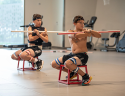
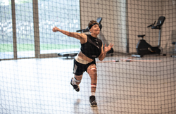
What if you could prevent a sports injury before it happens? That is exactly what the experts in the Center for Excellence in Sports Medicine at Scottish Rite for Children aim to achieve. Led by Sophia Ulman, Ph.D., the SAFE (Sports-specific Assessment and Functional Evaluation) program uses 3D motion capture technology — the same technology used to create special effects in movies and video games — to assess injury risk by studying the movements of young athletes.

The goal of the SAFE program is to develop injury prevention tools that will reduce sport-related injuries in young athletes here and beyond, creating a new standard of care. “Current injury prevention methods are falling short, as sports injuries in young athletes are skyrocketing,” Ulman says. “We are the first to use machine learning techniques to assess a young athlete’s risk for injury by considering risk factors such as movement characteristics, demographics, sports participation characteristics, psychological measures, nutrition, and sleep patterns.”

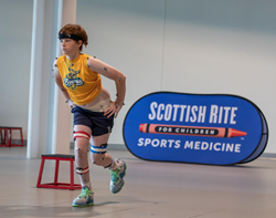
Uninjured athletes are invited to the Scottish Rite’s Movement Science Laboratory, where they are fitted with reflective markers. The markers are used to collect the athlete’s movement patterns to evaluate their mobility, speed, agility, and power in 3D. Participants are asked to jump, squat, run, pivot, and perform sport specific movements like penalty kicks, layups, or back handsprings. “The data is analyzed to learn more about how young athletes move and to determine if certain movement patterns lead to future sports injuries that would require treatment,” Ulman says.

The SAFE program has tested the movements of approximately 340 athletes across sports, such as baseball, basketball, gymnastics, softball, track, and more. “After initial testing, we follow athletes for one year and note if any athlete experiences an injury,” Ulman says. “This data is helping us compare the movements of athletes who remained healthy versus athletes who were subsequently injured to determine what may have led to the injury.”

Researchers at Scottish Rite have already identified findings that might help predict injury risk in young athletes. “We have found that current tools for assessing injuries of the knee may be unreliable in some instances,” Ulman says. “Current methods commonly rely on 2D video to assess injury risk, but our research using 3D modeling is identifying potential risk factors that the 2D assessment cannot.” Through this innovative program, Scottish Rite researchers collaborate with medical professionals and sports medicine experts across the field to advance the treatment of young athletes throughout the country.

The SAFE program is poised to change the way health care professionals assess athletes for injury risk and, best of all, will help keep young athletes in the game.

To learn more about Movement Science, please call 469-515-7160 or email MSL.Frisco@tsrh.org.

Read the full issue.

Osteochondritis Dissecans (OCD) in the Elbow

Osteochondritis Dissecans (OCD) in the Elbow

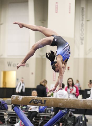
Our Center for Excellence in Sports Medicine treats a wide array of sport-related injuries and conditions in young athletes. One common condition treated is osteochondritis dissecans (OCD) of the elbow. This condition can happen to anyone but is especially common in sports such as gymnastics, tumbling, and baseball.

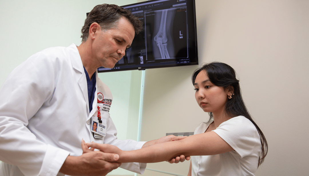
“This condition often presents to us in very late stages because it develops without symptoms,” says pediatric orthopedic surgeon Philip L. Wilson, M.D. He advises athletes, particularly baseball players and those in weightbearing sports like gymnastics, not to ignore nagging elbow pain. “Painless loss of extension is another sign that should not be ignored,” he says. “Proper diagnosis and early treatment can make a real difference in the course of care and outcomes.”

Our pediatric sports medicine team is a national leader in caring for and studying elbow OCD in young athletes. “The more we learn about the condition and the athletes, the better we can be at treating elbow OCD and teaching others the best way to prevent and manage it,” Wilson says. Here are two examples of Scottish Rite’s work:

  • An ongoing study called SAFE is open to young athletes, including gymnasts and baseball players. This study is looking at movement mechanics and the causes of injuries in these populations. Check out this video about SAFE testing.

  • study published in 2021, “Elbow Overuse Injuries in Pediatric Female Gymnastic Athletes: Comparative Findings and Outcomes in Radial Head Stress Fractures and Capitellar Osteochondritis Dissecans,” specifically addressed findings in 58 elbows in gymnasts (average 11 years of age) treated at Scottish Rite for Children throughout a course of five years. This study was the first to describe the differences between OCD and radial head stress fractures.

Learn more about OCD of the elbow, its causes, symptoms, treatment, and prevention below.

What is osteochondritis dissecans of the elbow?
The surfaces of the bones inside joints are covered with a smooth, gliding surface called cartilage. Osteochondritis dissecans (OCD) is a condition in which an area of cartilage and the underlying bone begin to soften, crack, or even separate. If left untreated, OCD can cause further damage to the cartilage in the joint and early arthritis.
This is a rare condition that most often affects the knee, but it can also affect the elbow, hip or ankle. In the elbow, the surface on the end of the humerus, the capitellum, is the most affected. This is typically seen in active individuals ages 8 to 19, more often boys than girls.

How does elbow OCD occur?
There are likely several factors, and the exact cause is still unclear. A common cause is a temporary loss in blood supply to an area of bone in a growing child, often combined with repetitive joint impact (overuse). There may be a genetic cause as well. Athletes at risk also often have a history of early sport specialization and year-round training. Some may report a history of a minor injury, but this is likely not the cause of the OCD lesion.

What are the signs and symptoms of OCD in the elbow?
OCD may be present even if there are not symptoms. An asymptomatic OCD lesion, one that does not cause any symptoms, may be identified when evaluating another concern. Signs and symptoms vary and may include:

  • Pain that worsens with activity
  • Popping or clicking
  • Swelling
  • Fluid inside the joint
  • Catching or locking with movement
  • Limited motion

How is elbow OCD diagnosed?
Physical examination, history, and X-rays are used to diagnose OCD in the elbow. Advanced imaging, such as an MRI, is often necessary to fully assess the condition and determine treatment options.

How is elbow OCD treated?
Properly treating and managing osteochondritis dissecans in the elbow lowers the risk of long-term damage to the joint. With diagnosis and treatment in the early stages, tissues may heal with rest and limiting activities that cause pressure on the OCD lesion.

Athletes benefit from continued training while resting their elbows. It is important for our team to help them understand what activities are safe and will not cause further problems on the elbow. Examples of activities to continue while receiving treatment for elbow OCD include:

  • Jogging
  • Stationary bike
  • Core strengthening
  • Lower body weightlifting of resistance training
  • Swimming
  • Golf putting only

These “weightbearing” activities are not allowed because they put pressure directly on the area of the OCD lesion:

  1. Sports of any kind
  2. Handstands
  3. Tumbling
  4. Push-ups, planks
  5. Upper body weightlifting or resistance training

When may surgery for elbow OCD be needed?
Many elbow OCD lesions can improve with conservative, nonoperative treatment. However, surgery may be necessary if the:

  • The OCD lesion appears loose, unstable, or large.
  • Cartilage becomes loose in the joint.
  • Imaging shows an advanced or worsening condition.
  • Symptoms are worsening despite nonsurgical treatment.

What kinds of procedures are used to treat OCD in the elbow?
The choice of surgical procedure depends on the condition of the tissues at the time of surgery. Most procedures are performed using an arthroscope, a camera, and tools inserted through small incisions, but a large surgery may be needed in some cases. Our sports medicine pediatric orthopedic surgeons are experts at treating OCD and can walk you through what to expect.

Procedures that may be offered alone or in combination include:

  • Drilling – drilling holes into the bone to increase blood flow and healing.
  • Stabilizing – inserting a screw, suture, or other piece of hardware to keep loose tissue in place.
  • Grafting – placing biological tissue in the area.

What can be expected after surgery for elbow OCD?
Our sports medicine experts work with every patient to develop an individualized postoperative treatment plan. After surgery, closely following postoperative instructions will protect the joint while the tissue is healing. Exercise and activity recommendations will be different for every patient.

How long does OCD in the elbow last?
Each case is unique, and the timing of returning to normal activity or sports will be discussed with your sports medicine physician, surgeon, or advanced practice provider. Symptoms may last months or years. It’s very important to understand that symptoms may return if the area does not fully recover before returning to repetitive or weight-bearing activities.

How can elbow OCD be prevented?
Overuse injuries like OCD occur with a high volume of training, repetition of certain movements, and early specialization in a sport.

These suggestions can help to prevent elbow OCD and other similar conditions:

  • Learn how to moderate training loads and intensities.
  • Make time for free play and lifetime sports like tennis, golf, cycling, and hiking.
  • Take breaks weekly and between seasons.
  • Learn to properly warm up and perform conditioning for your sport.

Learn more about sport specialization and preventing overuse injuries in young athletes.

Common Youth Ice Hockey Injuries and How to Avoid Them

Common Youth Ice Hockey Injuries and How to Avoid Them

Ice hockey is a contact sport that carries a higher risk of injury as the skill and competition level increase. Though ice hockey may not be as prevalent in North Texas as it is in other regions, Scottish Rite’s Sports Medicine team still takes care of injuries from this fast-paced and fast-growing sport.

Rules, such as delaying body checking, change an athlete’s risk of injury. We asked Jacob C. Jones, M.D., RMSK, and Madelyn White, P.T., D.P.T., to answer a few questions about pediatric sports medicine and physical therapy as it relates to ice hockey. Here’s what they had to say.

What do we know about ice hockey injuries?
Injuries occur quite frequently. Even though much of youth hockey prohibits checking and overt contact, that doesn’t entirely eliminate all contact in the sport. Both acute and chronic musculoskeletal injuries happen to hockey players. Concussions are also common in ice hockey players.

Are there different injuries in different age groups or skill levels?
Although many injuries are similar, younger hockey players may have more frequent injuries involving their growth plates since older adolescents may be near completion of growth. As a result, providers should be aware of how any injury may affect a growth plate. This can help provide the best treatment and avoid future complications.

Are there certain considerations when a player returns to the ice after an injury and rehabilitation?
It’s important to allow a gradual return to full participation in on-ice and off-ice training after injury. Be sure to warm up adequately prior to practice and games and avoid playing through pain.

Are ice hockey injuries preventable?
Yes, some of them may be preventable, especially the chronic injuries. Different youth leagues around the world have implemented rules changes regarding checking to help reduce the incidence of acute injuries.

What are tips for a skater to help prevent injuries?
Wearing proper gear, continuing to build flexibility and core strength. Some common injuries include ankle and shoulder injuries. Focusing regularly on exercises that help with the strength and mobility of these areas could help prevent injuries. Many overuse injuries and burnout can be prevented by trying to find at least three months out of the year to do a non-hockey sport or activity.

What should parents know about concussions in ice hockey?
Like other contact sports, these happen even with appropriate protective equipment and rules to avoid contact. Given the unique nature of the sport of hockey compared to other field sports, a treatment plan and return to play program should be tailored to hockey players. Seeing a medical provider familiar with sport-related concussions and hockey can help determine when it is safe to return to the ice, then safe to return to full hockey participation.

Are there exercises to help prevent back pain in ice hockey skaters?
Exercises to maintain hip mobility can help maintain good skating form and avoid back pain during hockey. It’s also important to build up abdominal and glutes strength. Exercises such as planks, banded side steps and crab walks can help prevent low back pain.

Concussion Balance Study

Concussion Balance Study

Learn how we use our Movement Science Lab to evaluate balance testing in sport-related concussion management.

Balance testing is commonly used to assess impairment and recovery after a sport-related concussion in the clinic setting. Measuring imbalances while going through various stances combined with both a firm and foam surface can provide valuable information in the evaluation of a concussion. Scottish Rite’s study, recently published in Brain Injury, was designed to look at how balance performance differed from diagnosis to return-to-play among athletes recovering from a concussion. A standardized test called the Balance Error Scoring System (BESS) is easy to perform in a clinic setting, but it may not provide the level of detail needed for a research study evaluating balance after sustaining a sport-related concussion. By conducting balance testing using the Movement Science Lab’s force plates, or special areas built into the flooring that are sensitive to the weight and force applied, researchers could correlate the BESS results with a highly objective center-of-pressure (COP) measure.

Principal investigator and director of movement science Sophia Ulman, Ph.D., explains the differences between these tests. “The BESS is a subjective test that requires clinical training and practice,” she says. “Alternatively, the force plate used to assess COP provides very specific, multidimensional measures that allow for discrimination of small differences in balance performance.”

It has been well established that there is an increased risk of prolonged symptoms as well as potential for compounding injuries if an athlete returns to play too soon after a sport-related concussion. Although balance is not the only measure used to determine readiness for sport, the proper assessment of balance is an important factor in this decision making. After reviewing data for these two tests in 40 patient-subjects, our team noted that the commonly used BESS test may not provide the information needed to assist with balance assessment as symptoms improved.

What does this mean for providers managing sport-related concussions?

Despite the volume of studies on the topic, the Sports Medicine team is continuing to learn about managing sport-related concussions in young and growing athletes. Pediatric sports medicine physician Shane M. Miller, M.D., says, “Until there is a better test to use in the clinical setting, we will continue to use tests like the BESS to do our best to assess balance improvement and identify the right time to return athletes to their sport. I suspect this will be a conversation for many years.”

The study, “Improvement in balance from diagnosis to return-to-play initiation following a sport-related concussion: BESS scores vs center-of-pressure measures,” was published in July 2022 in Brain Injury, the journal of the International Brain Injury Association.

Does my child need surgery to fix a clavicle fracture?

Does my child need surgery to fix a clavicle fracture?

Pediatric orthopedic surgeons Henry B. Ellis, M.D., and Philip L. Wilson, M.D., along with colleagues from the multicenter study group Factors Associated with Clavicle Treatment Study (FACTS) have published another set of findings in the American Journal of Sports Medicine. This group, like many others in pediatric orthopedics and sports medicine, merges the experiences and data from across institutions to provide the best evidence for care in the pediatric population. This group focuses their efforts on collarbone (clavicle) fractures and injuries in children and adolescents.

Here are some highlights from the publication. You can also visit the journal’s website to read the full article.

  • Midshaft clavicle fractures most often occur in adolescents, yet, most medical evidence is in adults until now.
  • More than 400 patients (10 to 18 years) with 100% displaced clavicle fractures were included in the study.
  • After two years, there was no difference in outcomes between those that had surgery and those that did not.
  • Those who underwent surgery had more nerve damage (loss of sensation on their chest wall) and more second surgery to remove plates and screws.
  • The study conclusion states, “Surgery demonstrated no benefit in patient-reported quality of life, satisfaction, shoulder-specific function or prevention of complications after completely displaced clavicle shaft fractures in adolescents at two years after injury.”

So, the answer to the question, “Does my child need surgery to fix a clavicle fracture?” is not yes. But, that also does not mean it is no. The study describes the general experience of a large group patients who have and have not had surgery for this condition. The individualized assessment of the patient is still important and necessary, but the study does show that there is not an obvious answer that applies to all patients. “This work is new and very important for the growing body of evidence in caring for this population,” Ellis says. “We can confidently tell families that one path is not yet obviously better than another.” In our individualized patient care, it is important for us to provide evidence-based recommendations, and in our research, we aim to define the recommendations.

This study, Two-Year Functional Outcomes of Operative vs Nonoperative Treatment of Completely Displaced Midshaft Clavicle Fractures in Adolescents: Results from the Prospective Multicenter FACTS Study Group, was published in the American Journal of Sports Medicine in September 2022.

Current Concepts: Management of Acute Shoulder Instability in Young Athletes

Watch the full lecture.

Our latest presentation from Coffee, Kids and Sports Medicine covers the management of acute shoulder instability in young athletes. Sports medicine physician assistant Ben Johnson, P.A.-C., dives into the differences in instability patterns between the skeletally immature and skeletally mature shoulder, on-field/acute management of shoulder dislocation and evidence-based recommendations for treatment of first-time shoulder dislocation.
 
Johnson begins the presentation by discussing the epidemiology of shoulder dislocations in high school and collegiate athletes in the United States and explains why it matters. He then shares important insight in how children and adolescents differ from adults physically, especially in relation to the capsular elasticity, a smaller anterior-inferior recess and more. Johnson shows the changes that occur in the shoulder as an adolescent enters puberty, and he discusses how this affects shoulder injuries.
 
Next up, Johnson teaches on-field and acute management of shoulder dislocations in pediatric patients and what medical providers need to know. Acute management includes taking a brief history, initial assessment and considering sport-specific factors. He breaks down when and how on-field reductions should be performed, sharing the benefits of early reductions and red flags to consider.
 
Johnson provides an evidence-based review of external and internal immobilization compliance and outcomes, along with the pros and cons of each method. He then answers questions about operative versus nonoperative treatment, breaking down the consequences of each.
 
To wrap up the presentation, Johnson provides a summary on pediatric glenohumeral dislocation and the steps that should be taken when assessing treatment strategies, as well as sharing a treatment algorithm. The presentation is crucial for sports medicine physicians and other medical professionals who treat young athletes, especially those at a high risk for shoulder injuries.