Four Tips to Prevent Injuries in Youth Lacrosse

Four Tips to Prevent Injuries in Youth Lacrosse

Lacrosse is one of the oldest sports in North America and is also one of the fastest growing sports in the United States. In 2021, there were more than 40,000 collegiate and more than 450,000 youth lacrosse players. Boys’ and girls’ lacrosse follow different rules and require different equipment, which may impact the types of injuries seen in these young athletes.

“Lacrosse is an intense and demanding sport,” orthopedic surgeon John E. Arvesen, M.D., says. “Athletes who are prepared can dramatically reduce the risk of injury.” Coaches and parents can use these tips to guide young athletes in the right direction.

FOUR WAYS TO REDUCE INJURY RISK IN LACROSSE

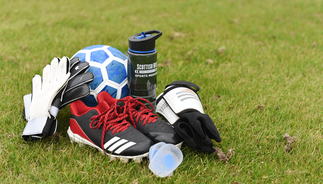
Wear proper equipment. Protective gear that meets standards and fits correctly will provide the most benefit. Poor-fitting equipment may not offer the same protection or support.

Perform a dynamic warm-up. This involves continuous movement to raise the body’s core temperature in preparation for training or competition. Perform this before each practice or game to increase elasticity in the muscles, tendons and ligaments around the joints.

Learn and implement effective hydration strategies. Mild dehydration can worsen performance. Ideally, players should drink water every 15 to 20 minutes. Add a sports drink during intense activities lasting longer than one hour or in very hot environments to replace electrolytes lost through sweat.

Plan for rest and cross-training. Early sport specialization increases an athlete’s risk of injury and inhibits their athletic development. Focusing on one sport at an early age may lead to movement imbalances, an increased risk of injury and overtraining. A one- or two-month break between seasons and a day or two of rest each week in-season is recommended.

Some injuries in lacrosse are more difficult to prevent, such as those that occur from sticks, collisions and falls. Non-contact injuries are considered to be more preventable. As a sports physical therapist and youth lacrosse parent and coach, Michael Losito teaches young athletes the importance of learning fundamental movements to prepare their bodies for non-contact injuries. “When an athlete has control over the trunk and lower extremities, he or she can produce more power, which may help to protect the joints from minor and serious injuries,” Losito says.

Common Injuries in Lacrosse

Despite efforts to reduce the risk of injury, some accidents are going to happen. Make sure you recognize and respond to injuries promptly. Many conditions can be treated with rest and a short round of rehabilitation if they are recognized early.

SPORT-RELATED CONCUSSIONS
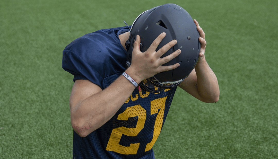
Concussions in girls’ lacrosse players are often the result of stick contact, or a blow with a stick to the head. Boys’ lacrosse has a higher risk for concussion due to player-to-player contact. An athlete with any concussion symptoms, including change in behavior, thinking or performance after a collision or a blow to the head, neck or body, should be removed from play immediately and not return to play the same day. A medical professional with experience managing concussions should determine when it is safe to return to play.

KNEE AND ANKLE INJURIES
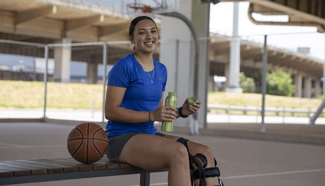
Sudden changes in direction, stopping and jumping can place a lot of stress on the knee.  This can stretch and tear ligaments, such as the anterior cruciate ligament (ACL) and medial collateral ligament (MCL). A swollen knee is a sign of a joint injury that needs to be evaluated.

In lacrosse, sudden direction changes, stepping on another player’s foot or landing from a jump can result in an ankle injury. Ankle sprains and injuries to the growth plate are common in growing athletes and should be evaluated to optimize treatment and return to sport.

BACK AND CHEST INJURIES
Player-to-player collisions or falls may cause back injuries. Powerful and repetitive rotation while running, cradling, shooting and passing is more likely to cause activity-related pain in lacrosse. Overuse injuries, such as stress fractures (spondylolysis), are also common in young athletes. Persistent back pain from overuse injuries needs to be evaluated by a medical professional. The equipment and high-speed movements in lacrosse increase the risk of a rare injury from a direct blow to the chest from the ball, a stick or player collisions. The condition, called commotio cordis, can be life-threatening. Chest protectors may reduce the impact and risk of this injury.

ARM INJURIES
The design of lacrosse protective gear allows the arm to move freely but leaves the shoulder open to injury. Clavicle (collar bone) fractures and ligament injuries, or “separated shoulders,” may occur. A change in the appearance of the shoulder, pain, swelling or limited motion after a collision or fall should be evaluated.
Body checks, stick checks and slashing may cause hand and wrist injuries. Soft tissue injuries such as ligament sprains may heal with rest and ice. Others may need a brace or other treatment.

Download the PDF.

Does a Discoid Meniscus Injury Need Surgery?

Does a Discoid Meniscus Injury Need Surgery?

A discoid meniscus is an abnormally shaped piece of cartilage found in the knee joint, and due to its shape, twisting knee movements can sometimes cause it to tear. When determining whether treatment for this injury is necessary, it is important to consider why, when and how the condition was discovered.

What is a meniscus?
The round end of the femur (thigh bone) sits on the flat top of the tibia (shin bone) to make up the knee joint. The femur is supported by the meniscus, which is composed of two soft “c” shaped cartilage structures. They act like soft cushions that help support the knee joint. The one that sits on the inner side is called the medial meniscus, and the one on the outer side is called the lateral meniscus.

What is a discoid meniscus?
Instead of having the typical “c” shape, a discoid meniscus forms as a solid piece, like a disc or a Frisbee®. The tissue grows thicker and larger than a normal meniscus and also has an abnormal texture, which makes it more likely to cause problems.

What causes a discoid meniscus?
A discoid meniscus is a congenital (at birth) defect and does not grow into the normal shape. This defect is not caused by trauma (i.e., an accident) or an injury. One to two out of every 100 children have a discoid meniscus. The condition is found more often in boys.

A discoid meniscus cannot be prevented. As the child grows, injuries and/or changes in the alignment of the hip, knee and ankle may cause symptoms.

What are the symptoms?
A discoid meniscus does not always cause symptoms. It may go unnoticed until symptoms begin. Symptoms can include pain, popping or snapping, limping, inability to bear weight (stand or walk) and inability to straighten the knee.

How is a discoid meniscus diagnosed?
A thorough history and physical examination are used to diagnose a discoid meniscus. Common findings on the outside of the knee (lateral joint line) include a bulge that can be seen or a “snap” that can be felt and heard.
X-rays are used to look at the alignment of the bones in the knee and leg. Other imaging, such as an MRI, may be used to look at the condition of the meniscus and other tissues in the knee.

What is the treatment?
For children who do not have symptoms or if they have a “clunk” when they move their knee, yet do not experience pain or difficulty conducting daily activities, no treatment is needed.

Early symptoms, such as swelling and pain, can be managed by resting, elevating the leg and other common strategies for knee injuries, such as ice and anti-inflammatory medications.

Surgical treatment is needed if there is a concern regarding the development of the knee with a large discoid or when symptoms begin to interrupt daily activities.

A knee arthroscopy, a type of minimally invasive surgery, may be recommended. The goal of surgery is to improve the shape of the meniscus and remove any loose or extra tissue that may cause the joint to become stuck. Rehabilitation and a slow return to sports may be necessary after surgery to change the shape of the meniscus.

A discoid meniscus increases the risk of a meniscal tear, and therefore, the condition is often found when evaluating an MRI of the knee after an injury. In these cases, treatment may be recommended to improve the shape of the meniscus. This can be done at the same time as surgery for other problems diagnosed in the knee.
What is the long-term outlook?

A discoid meniscus should not prevent normal daily activities or participation in sports. Diagnosis and management of symptoms can reduce the risk of further damage in the knee joint and prevent long-term problems. Regular follow-up to monitor the growth and health of the developing joint is very important after diagnosis, even if treatment is not needed in the early stages.

An important initiative of the Center for Excellence in Sports Medicine team at Scottish Rite for Children is a quality improvement registry designed to learn about the care and outcomes of treatment for discoid meniscus, among other conditions. This multi-center collection of data is led by pediatric orthopedic surgeon and director of clinical research Henry B. Ellis, M.D., is called the Sports Cohort Outcomes Registry (SCORE).

“This large collection of data allows us to compare surgery findings and outcomes across different age groups. The data set is unlike any other and will help to define care for this condition and many others. Early results were shared at the Pediatric Research in Sports Medicine annual meeting in 2022 and have already shaped more studies and better patient care.”        
– Henry B. Ellis, M.D.

Each institution in the SCORE group may take care of a handful of patients with this condition each year. The compiled data, reviewing nearly 300 patients and their outcomes helps to provide better education to patient-families, improve surgical decision-making and setting better expectations for outcomes.

Differences in the appearance of the meniscus as well as the ability for the meniscus to be repaired were apparent. In younger patients, the meniscus:

  • Is larger and covers more of the bone.
  • May have loose, unstable edges.
  • Is more likely able to be repaired.

These early findings help pediatric orthopedic surgeons know what to expect and how to counsel parents about who may or may not need surgery. Ultimately, the registry will be able to provide standard outcome expectations which will further improve the patient experience and outcomes.

Acute Ankle Injuries in Youth Sports

Acute Ankle Injuries in Youth Sports

This is a summary of a program presented as part of a free, monthly education series at Scottish Rite for Children in Frisco, Texas.

Register for this and other on-demand programs or watch the presentation on our YouTube channel for Medical Professionals.

Download a PDF of this summary.

Shane M. Miller, M.D., discussed commonly encountered acute ankle injuries in a young athlete including considerations for safe return to play after an ankle injury.
Ankle injuries are unfortunately very com­mon in young ath­letes, studies mentioned estimate:

  • an­kle sprains ac­count­ for 16% of all in­juries.
  • ankle injuries represent 22% to 50% of all sport-related injuries presenting to emergency departments.
  • one in four of all recurrent injuries among high school athletes are in the ankle.

Additional factors noted about the epidemiology of ankle injuries include:

  • Girls have a higher injury rate than boys in soccer, softball/​baseball, and track and field, but similar rates are observed in basketball, volleyball and lacrosse.
  • Dynamic sports requiring jumping and cutting activities, such as basketball, gymnastics, volleyball, soccer and football account for the majority of acute ankle injuries.
  • Indoor court sports and sports that involve player-to-player contact are high risk.
  • Sports involving repetitive activities and running, such as cross country, track and field, gymnastics and soccer, are commonly associated with overuse injuries of the ankle.

History and Evaluation

When discussing the athlete’s history, elements to consider include variables beyond age, sex and sport. Knowing the position played, level of competition and history of previous injuries (same side, opposite side, knee, concussion, etc.) will help in understanding the conditions surrounding the injury. Understanding the long-term goals of the athlete and timing (pre-season, playoffs, etc.) helps to customize the treatment planning and anticipate the athlete’s response to the plan.
When assessing the history of the injury, ask:

  • Is this the first time you have had any pain or instability in your ankle? (Acute or Chronic?)
  • How did the injury occur?
  • Was there any swelling?
  • Did you hear or feel a pop?
  • Were you able to walk on it?
  • Were you able to keep playing?
  • Can you point with one finger to the location of pain?
  • What treatment was provided immediately after it occurred and since that time?

Commonly encountered acute ankle injuries in a young athlete

Using a case-based approach, Miller covered common acute ankle injuries and approaches to evaluation and early management. He reviewed key elements of each case using these questions as a guide:

  • What is the most likely diagnosis and mechanism of injury?
  • When is imaging necessary and what would you order?
  • What does your initial treatment entail?
  • When should referral to an orthopedic/sports medicine specialist be made?

Ankle Sprain

A strain is a muscle injury. A sprain is a ligamentous injury, and most, approximately 85%, are inversion injuries and involve the anterior tibial fibular ligament (ATFL) (lateral ankle sprain). Injuries to this and other ligaments are commonly associated with bruising, swelling, inability to bear weight and limited range of motion. These injuries also tend to have a high rate of recurrence or chronic instability.

Imaging

With the presence of bony tenderness, inability to bear weight or significant swelling or bruising, anterior posterior (AP), lateral and mortise views are recommended. Ottawa ankle rules are helpful in determining if X-rays of the ankle are necessary in adults and children. Tenderness in other areas may indicate additional X-rays of the foot. Because an MRI is rarely needed, a specialty referral is indicated if considering an MRI for an ankle sprain. An MRI may be helpful to evaluate for some conditions like occult fractures or more significant injury, such as high ankle sprains or an osteochondral injury.

Treatment

Early treatment of acute injuries of the ankle should include strategies that protect the ankle from further injury, reduce and prevent swelling and promote early mobilization. A familiar pneumonic, “RICE” has been modified to, “PRICEMMS” to include treatment beyond the early acute stage.

  • Protection from further injury – walking boot, splint, ankle brace or air stirrup
  • Relative Rest – not doing anything that hurts, including the use of crutches if the patient is limping.
  • Ice – 20 minutes on the hour may help with pain and to reduce swelling
  • Compression – ankle wrap
  • Elevation – above the level of the heart
  • Medications – analgesics and anti-inflammatories
  • Mobilization – improving range of motion using gentle, early mobilization including active dorsiflexion and multi-directional movements, i.e., making letters of the alphabet with the toes.
  • Strength – training of the peroneal and gastrocnemius muscles with high repetition, isometric and low resistance exercises, balance exercises and proprioception training

Return to play after an ankle sprain

In general, young athletes with musculoskeletal and cervical spine injuries should not return to play until they have full range of motion, resolution of pain, normal strength, psychological readiness and the ability to demonstrate adequate sport-specific skills.
Return to play criteria should include:

  • Absent (or minimal) pain
  • Stable ankle with daily activity
  • Full range of motion
  • Normal strength (>90% of uninjured side)
  • Good balance/proprioception
  • Able to protect self from further injury
  • Functional progression – maneuvers at full speed, no pain
  • Restoration of confidence

After considering these items, individual circumstances should be assessed to identify risk of further injury and the need for protective bracing or additional time or treatment prior to returning. In some conditions, a referral to a pediatric sports or orthopedic provider may be advised, these include:

  • Confirmed or suspicion for fracture
  • Syndesmosis or “high ankle” sprain
    • Damage to the anteroinferior tibiofibular ligament (AITFL) and syndesmosis
    • Stress in external rotation and dorsiflexion will exacerbate pain.
    • May see widening of mortise on X-ray
  • Recurrent ankle injuries
  • Need for rapid return to sports participation
  • Not responding to normal conservative treatment

Physeal Injuries

Physes, commonly referred to as growth plates, are composed primarily of cartilage cells so are more susceptible to both acute and overuse injury. The physis is the “weak link” in the chain and injuries in this area may lead to growth arrest or deformity.
Key terms to know:

  • Diaphysis – midshaft, tubular portion of long bone
  • Metaphysis – area adjacent to physis, consists of cancellous bone
  • Physis – growth plate
  • Epiphysis – longitudinal growth center
  • Apophysis – growth center that adds contour to a bone

Often a site of muscle/tendon attachment

Ankle Physeal Injuries

Salter-Harris Fracture Classification​

  • I and II – don’t involve joint surface, usually do well without surgery
  • III, IV and V – involve articular surface, need specialist consultation
  • Salter-Harris I – must have high index of suspicion
    • X-rays may be negative with Salter-Harris I
    • Comparison views may be helpful
    • However, they may be less likely than previously thought
    • Boot may be preferred over a cast, when immobilization is indicated
  • Radiographic stress views are discouraged.
  • Beware of the medial ankle sprain—tibial physeal injuries are a more likely diagnosis.
  • Occult fractures can cause gait disturbances in young children.
  • An ankle injury in a prepubertal adolescent may be a growth plate fracture rather than an ankle sprain.
  • Presence of a subfibular ossicle may be related to a prior ankle injury, but treatment is not indicated unless it is symptomatic.
  • Transitional fractures include triplane and Tillaux fractures.
    • These occur as the growth plate is closing.
    • These typically need surgical intervention.

Prevention

Studies reviewed in this presentation compared types of off-the-shelf ankle braces. Results suggest that braces may reduce the incidence but not reduce the severity of ankle, knee or other lower extremity injuries. Balance training was a finding in an article reviewing lateral ankle injury studies. Co-course director and program moderator, Henry B. Ellis, M.D., contributed to this review and provided comments in the Q and A session.

Key Takeaways

  • Foot and ankle injuries are very common in young athletes.
  • Have a high index of suspicion for fracture and low threshold to obtain X-rays.
  • Consider bracing and balance training for prevention of ankle injuries.

Five Signs Your Young Athlete May Be Underfueling

Five Signs Your Young Athlete May Be Underfueling

What is underfueling?

When athletes do not eat enough calories (or the right calories) to support their growth and development and all of the training. This may occur periodically due to fluctuations in seasons or training schedules, it is particularly concerning if it happens frequently.

How do I know if my athlete is underfueling?

Without complex calculations, parents can watch an athlete for signs and listen for complaints that indicate underfueling. Below is a short list to help.

  1. The athlete is no longer making improvements in skill and performance or has experienced a sudden decrease in performance.
  2. Injuries take a long time to heal or there are recurring injuries, like stress fractures.
  3. Weight loss that is not otherwise explained. It is important to consider that an athlete may not be losing weight but may still not be getting enough calories.
  4. Delayed growth and development. For a female athlete, an obvious sign is irregular or missed periods or a delay in starting her period.
  5. Frequent dizziness and headaches.
  6. Complaints of constant fatigue.

What should I do if I think my young athlete is underfueling?

  • Make sure he/she is getting three balanced meals a day. Busy teens tend to skip the breakfast meal.
  • Add one or two snacks a day. Often the most reliable snack to add is a bedtime snack. Elite young athletes, especially those struggling with underfueling, typically need at least 3 snacks a day.
  • Increase portions in current meals. Small increases throughout the day can make a difference.
  • Make nutrient-dense swaps in meals and snacks. This way your athlete doesn’t have to worry about adding more food or more eating instances in an already packed schedule.
  • Make drinks count. When athletes need extra calories, include beverages like milk, chocolate milk and calcium-fortified orange juice with meals. Offer sports drinks with practices.

Where can I turn for help if I am concerned?

If your child has lingering or recurrent injuries in sports, delay or changes in menstruation or other concerns that might be related to underfueling, our Sports Medicine team can help. After a medical evaluation with a sports medicine physician, additional services such as a consultation with a certified sports dietitian may be recommended. Call 469-515-7100 to request an appointment.  

Find more resources about sports nutrition for young athletes. 

Five Signs Your Young Athlete May Be Underfueling

Five Signs Your Young Athlete May Be Underfueling

What is underfueling?
When athletes do not eat enough calories (or the right calories) to support their growth and development and all of the training. This may occur periodically due to fluctuations in seasons or training schedules, it is particularly concerning if it happens frequently.

How do I know if my athlete is underfueling?
Without complex calculations, parents can watch an athlete for signs and listen for complaints that indicate underfueling. Below is a short list to help.

  1. The athlete is no longer making improvements in skill and performance or has experienced a sudden decrease in performance.
  2. Injuries take a long time to heal or there are recurring injuries, like stress fractures.
  3. Weight loss that is not otherwise explained. It is important to consider that an athlete may not be losing weight but may still not be getting enough calories.
  4. Delayed growth and development. For a female athlete, an obvious sign is irregular or missed periods or a delay in starting her period.
  5. Frequent dizziness and headaches.
  6. Complaints of constant fatigue.

What should I do if I think my young athlete is underfueling?

  • Make sure he/she is getting three balanced meals a day. Busy teens tend to skip the breakfast meal.
  • Add one or two snacks a day. Often the most reliable snack to add is a bedtime snack. Elite young athletes, especially those struggling with underfueling, typically need at least 3 snacks a day.
  • Increase portions in current meals. Small increases throughout the day can make a difference.
  • Make nutrient-dense swaps in meals and snacks. This way your athlete doesn’t have to worry about adding more food or more eating instances in an already packed schedule.
  • Make drinks count. When athletes need extra calories, include beverages like milk, chocolate milk and calcium-fortified orange juice with meals. Offer sports drinks with practices.

Where can I turn for help if I am concerned?
If your child has lingering or recurrent injuries in sports, delay or changes in menstruation or other concerns that might be related to underfueling, our Sports Medicine team can help. After a medical evaluation with a sports medicine physician, additional services such as a consultation with a certified sports dietitian may be recommended. Call 469-515-7100 to request an appointment.  

Find more resources about sports nutrition for young athletes. 

Scottish Rite For Children Adds ACGME-Accredited Sports Medicine Fellowship Program

Scottish Rite For Children Adds ACGME-Accredited Sports Medicine Fellowship Program

Scottish Rite for Children and UT Southwestern Medical Cente are beginning a new Accreditation Council for Graduate Medical Education (ACGME-accredited Sports Medicine fellowship program with a specialized pediatric focus. Led by program director Henry B. Ellis, Jr., M.D, and associate program director Philip L. Wilson, M.D, the program will take place primarily at the Scottish Rite for Children Orthopedic & Sports Medicine Center in Frisco. The first in the North Texas region, this new program is one of a small number of subspecialty training programs that provides significant exposure to the care of young and growing athletes

The field of pediatric sports medicine includes operative and nonoperative management of sport-related injuries and consideration of how conditions and treatment affect long-term athletic development during continued physical and mental maturation. The program provides in-depth training for managing common and complex conditions, including ACL injuries, osteochondritis dissecans, patellar instability, meniscus, hip preservation, shoulder instability and more. In the United States, approximately 45 million children between the ages of 5 and 18 participate in organized sports according to a paper published in theInternational Journal of Sport Communication, which makes the need for specially trained pediatric sports medicine physicians and surgeons important for the well-being of today’s youth

Fellows are frontline members of the sports medicine care team at Scottish Rite for Children Orthopedic and Sports Medicine Center in Frisco, Texas. In addition, they rotate with sports medicine surgeons at UT Southwestern and other adult colleagues for the full gamut of operative sports training. Additionally, the fellow assists in the management of pediatric fractures and acute orthopedic conditions at Children’s Medical Center Dallas, one of Texas’ only Level 1 pediatric trauma centers

The one-year fellowship program is available to postgraduate surgeons who have completed an orthopedic residency. The fellowship provides the recipient the opportunity to pursue advanced study, in-depth training, management and research in sports medicine, musculoskeletal, and complex injuries. The fellowship accommodates one fellow and begins on August 1

Learn more on our Fellowship & Graduate programs page