Nico is Spreading His Wings!

Nico is Spreading His Wings!

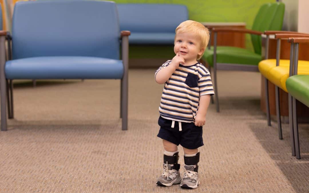
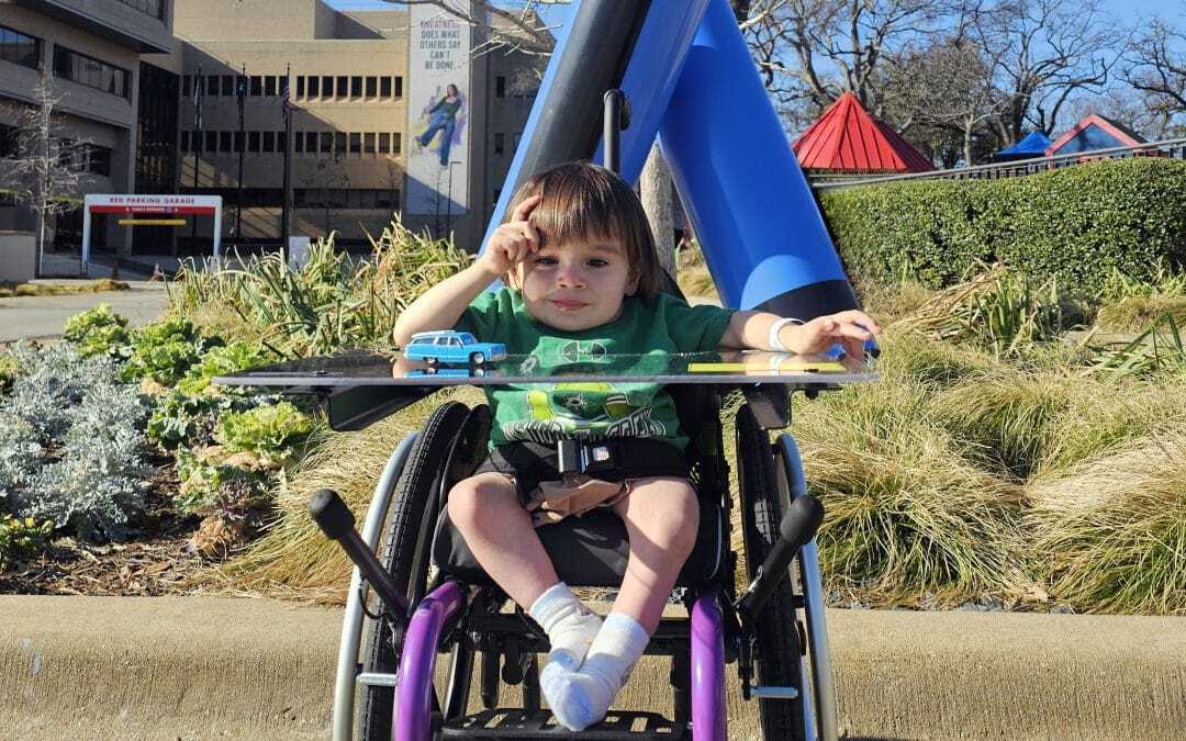
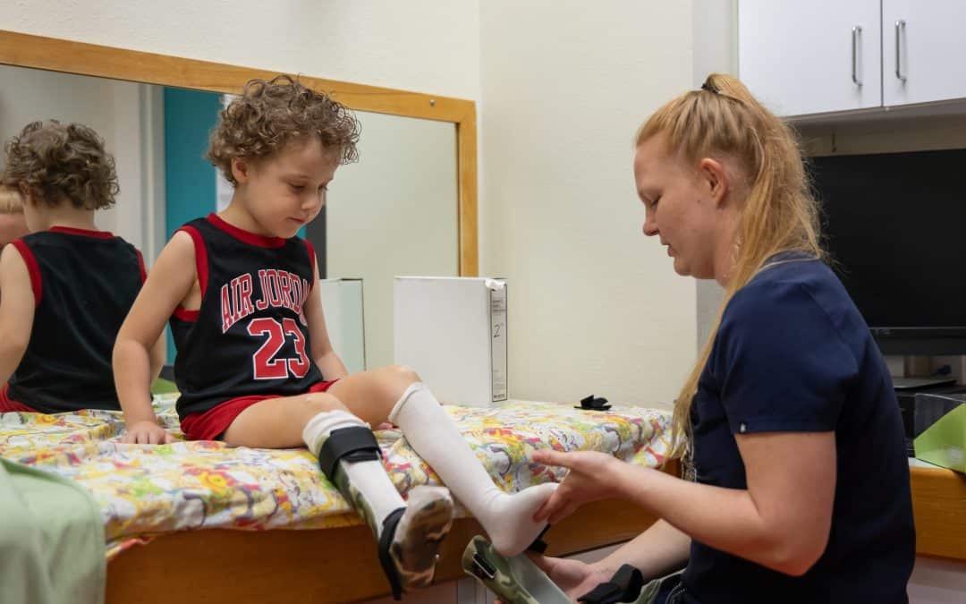
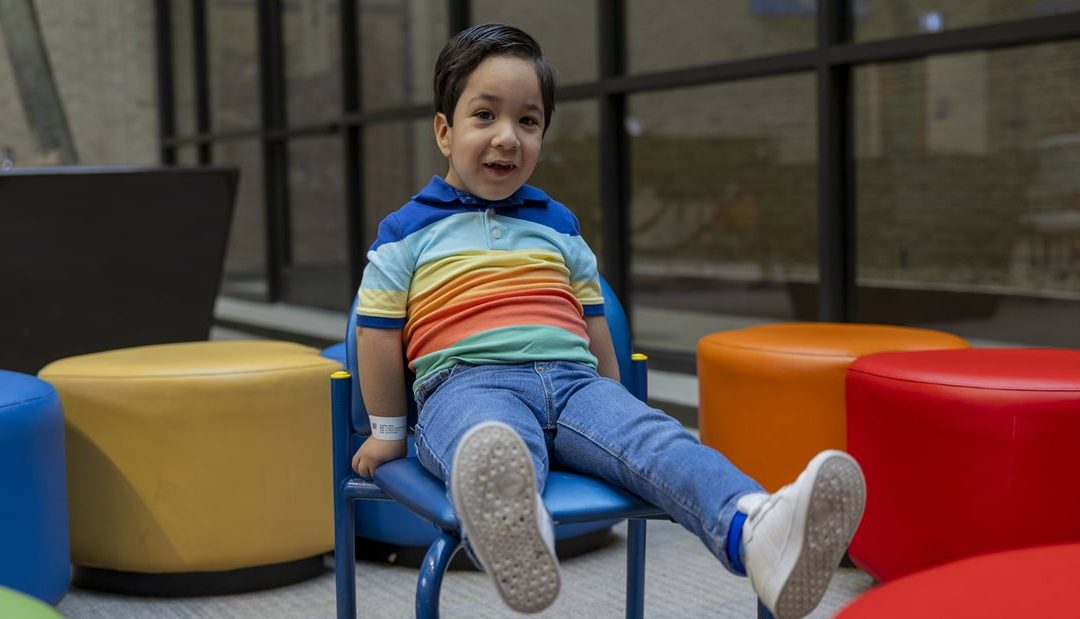
3-year-old Nico, of Mission, and his family travel more than 500 miles to receive treatment at Scottish Rite for Children. “We make that effort to come this far, because it’s worth our drive,” Nico’s mom, Mary says. Nico was diagnosed at birth with clubfoot and arthrogryposis — a rare condition that causes stiff joints. Mary found comfort in the arthrogryposis support groups she found online. Her conversations with other families led her to schedule an appointment for Nico at Scottish Rite.

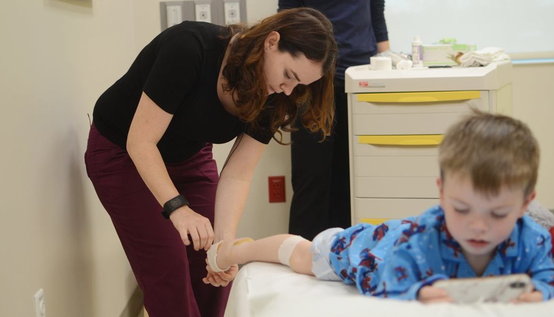
Nico receives care from our experts in hand, clubfoot, occupational therapy and orthotics. Even when he’s facing a challenge, Nico’s joyful personality shines. He is always greeting and waving at everyone he meets at Scottish Rite. “I think Nico is going to grow up to be an entertainer,” Mary says. “He is such a social butterfly!” 
When surgery was proposed as part of Nico’s treatment plan, Mary felt assured. “We were always confident about surgery,” Mary says. “The outcomes our care team told us were always what we received.” Nico has had two surgeries to combat his arthrogryposis: an Achilles lengthening surgery on both legs in 2022 and a wrist surgery in 2023. 

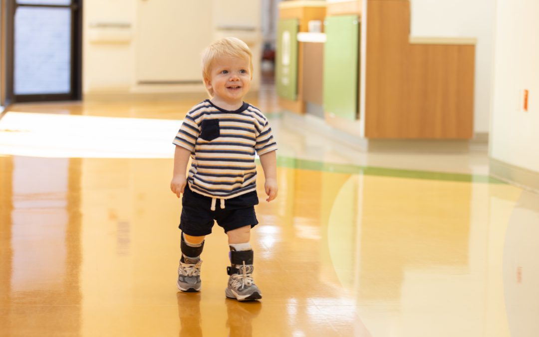
Nico has made remarkable progress, and there is no stopping him now! Mary says she is thankful to Dr. Scott Oishi and Dr. Anthony Riccio for their attentive care. With their help, Nico can now walk by himself. He’s also working with occupational therapists Amy Sitabkhan and Grace Evansco to develop more independence by learning daily tasks such as eating and clothing himself. “I wish I could take Scottish Rite’s occupational therapists back home with us,” Mary says. “Everyone is so helpful, and we are so blessed.”

We like to say all our patients become part of the Scottish Rite family. For Nico, this is especially true. He and his family are now part of our Hand in Hand support group, and it’s been so fun to watch Nico come out of his shell. We look forward to seeing everything he accomplishes in the future!


Do you have a story? We want to hear it! Share your story with us. 

What to Do When Your Child Has Pigeon Toes

What to Do When Your Child Has Pigeon Toes

Most feet point forward, aiming at their destination — but not all. Although this condition can cause concern for parents, intoeing, or pigeon toes, is common. Intoeing occurs when children have feet turned inward toward one another. Here’s what you should know to help your child manage this condition and enjoy an active and healthy life.

Why Children Experience Pigeon Toes

Pigeon toes can exist at birth but often become noticeable later in childhood. There are three main causes of intoeing, all of which usually resolve without treatment at different times in a child’s growth. These causes include: 

  • Metatarsus adductus. While in the womb, your baby’s position may force the front of the foot to turn inward. This position can result in metatarsus adductus, a condition in which the toes point inward while the outer edge of the foot is curved outward. Metatarsus adductus is a common cause of pigeon toes in newborns and infants. In rare cases, the condition can cause the foot to look like clubfoot. This condition usually resolves spontaneously by age 2.
  • Internal tibial torsion. In this condition, your child’s tibia, or shinbone, twists and turns the leg inward. This is also related to “packaging” of the legs in an inwardly twisted direction while in the womb. This condition usually resolves spontaneously by age 6.
  • Femoral Anteversion. With femoral anteversion, your child’s thigh bone, the femur, turns inward, causing the feet and toes to point inward as well. This condition usually resolves spontaneously by age 9. 

Intoeing often runs in families. Because of its genetic root, preventing pigeon toes is not possible. 

When to See a Provider

Thankfully, being pigeon-toed does not cause pain, and most infants and children grow out of the condition without requiring treatment. The bones correct themselves as the child grows older. However, some cases of intoeing do not resolve on their own and require medical care. 

Your child’s provider may refer you to a pediatric orthopedic specialist if:

  • Your child’s intoeing is accompanied by other foot abnormalities.
  • The foot cannot straighten out easily and feels stiff.
  • The intoeing causes pain, limping, swelling or difficulty walking.

How to Treat Pigeon Toes

The vast majority of children with intoeing due to any of the above causes will correct their rotational differences without any treatment whatsoever. When intoeing is severe and has persisted beyond the expected age at which the condition causing the intoeing should have resolved, treatment may be considered.

Your child’s provider will consider several factors when determining the most appropriate treatment. These include the cause and severity of the intoeing, your child’s age and how the problem affects your child’s daily life.

Based on this information, your provider may recommend one or more of the following pigeon toe treatments:

  • Serial Casting. A cast on your child’s leg and foot can, over time, correct metatarsus adductus. Your provider may use a series of casts as your child’s treatment progresses to continue straightening the foot.
  • Stretching. Babies with metatarsus adductus may benefit from gentle stretching. Parents can perform this physical therapy technique at home. If appropriate, your provider will show you how to stretch the foot in a way that encourages it to straighten out. 
  • Surgery. Older children with severe symptoms that don’t respond to other treatments may require surgery. During surgery, an orthopedic surgeon cuts the twisted bone and resets it in the proper position. Surgery is typically for children older than 9 who have difficulty walking due to intoeing.

You may see advertisements for special shoes or exercise programs designed to address the conditions that cause intoeing. However, the American Academy of Orthopedic Surgeons has stated these are not effective. Your best resource for help with pigeon toes is your child’s provider or orthopedic specialist. 

If you are concerned about intoeing, schedule an appointment with a foot conditions specialist at Scottish Rite for Children.

Clubfoot Treatment: What Are Your Child’s Options?

Clubfoot Treatment: What Are Your Child’s Options?

If your baby is diagnosed with clubfoot, your first question may be, “What can we do?” Fortunately, effective nonsurgical clubfoot treatment options are available, and most children don’t need an operation. A pediatric orthopedic surgeon can help you decide what treatment makes the most sense for your child.

The Basics of Clubfoot Treatment

One of the most common foot-related deformities that may be present at birth, clubfoot occurs when your child has an inward-turned, high-arched foot. It isn’t painful for your baby, but if left untreated, the condition can lead to foot pain in the future. In addition, untreated clubfoot can cause an abnormal gait and restrict activities later in childhood.

Usually beginning soon after birth, clubfoot treatment can provide correction to the condition, so your child’s foot will have normal function, and your child will be able to walk without pain. With a pediatric orthopedic surgeon’s help and your commitment to following the treatment plan, clubfoot doesn’t have to be a permanent deformity. Treatment can help prepare your baby to walk, run, jump and skip through childhood.

The Ponseti Method: The Gold Standard of Nonsurgical Clubfoot Treatment

For many babies, correcting clubfoot involves a multi-step treatment called the Ponseti method, which is named after the orthopedic surgeon who pioneered it. This treatment starts with stretching and casting.

Over four to five weeks, your child will see a pediatric orthopedic surgeon for weekly manipulations. This gentle stretching gradually moves the foot into the correct position. After each session, the surgeon will apply a full-leg Ponseti cast to help the foot maintain its position.

A tight Achilles tendon contributes to clubfoot, and most patients receiving the Ponseti method have an Achilles tenotomy procedure. This procedure is quick and simple, is done in the clinic and is not major surgery.

With local anesthesia numbing the back of your child’s ankle, his or her orthopedic surgeon will cut the Achilles tendon to loosen it. The cut is so small and precise that it won’t require stitches. Afterward, your baby will wear a cast for three weeks while the tendon heals to the correct length.

Clubfoot can return, even with treatment. Your child will have to wear a brace with a metal bar attached to special shoes for four years. He or she will start out wearing the brace 23 hours a day. Once your baby begins pulling to stand the brace is used during nighttime hours only until it’s time to stop bracing.

The French Method of Clubfoot Correction

The Ponseti method isn’t right for every child. In some cases, another nonsurgical clubfoot treatment, the French method, may be a better fit. For this treatment, you’ll work closely with your child’s orthopedic surgeon and a physical therapist.

  • The physical therapist will teach you how to gently manipulate, tape and splint your child’s foot, which, over time, moves it back into the correct position. You’ll repeat this routine daily for three months.
  • After three months, your baby will likely need to have an Achilles tenotomy to release the tendon.
  • Following the tenotomy, you’ll resume manipulating, taping and splinting the foot each day until your child reaches age 2 or 3. Your pediatric orthopedic surgeon will monitor your child’s progress and let you know when you can stop the daily regimen.

Treating clubfoot takes dedication, but the payoff is worth it. Your effort can set your child up for a lifetime of activity without foot pain.

Wondering what’s next for your baby after a clubfoot diagnosis? Schedule an appointment with a foot and ankle specialist who can recommend a treatment plan.

Ankle-Foot Orthosis Devices: A Guide for Parents

Ankle-Foot Orthosis Devices: A Guide for Parents

Health conditions that limit your child’s ability to walk, run, play and just be a kid can take a lot of fun out of childhood. An ankle-foot orthosis could be the solution your child needs, particularly if he or she has muscle or joint problems in the legs, ankles or feet. These specialized orthotics can help relieve uncomfortable symptoms, allowing children to return to their daily activities. 

What Is an Ankle-Foot Orthosis?

Ankle-foot orthoses (AFOs), sometimes called AFO braces, are devices designed to support and improve the function of your child’s foot and ankle. Pediatric orthopedic specialists often prescribe them to children with conditions that cause joint instability, irregular walking patterns and muscle weakness or stiffness. 

Your child might need an AFO if he or she has: 

·       Deformities of the foot or ankle

·       Fractures

·       Neuromuscular disorders, such as cerebral palsy, muscular dystrophy and spina bifida

·       Peripheral nerve injuries 

·       Spinal cord injuries

AFOs can also treat ankle instability caused by cerebral palsy and other neuromuscular disorders, congenital abnormalities and injuries. A child with ankle instability may have a hard time maintaining balance, walking or playing. An AFO provides stability, improves gait patterns and prevents additional complications associated with ankle instability.

Three Primary Types of AFOs

Typically made from lightweight and durable materials, such as plastic or carbon fiber, an AFO holds your child’s foot and ankle in the proper position. They can be custom made to fit your child to ensure a secure fit.

Many people confuse orthotics with prosthetics, but they are not the same. A prosthetic device replaces a missing limb, whereas orthoses and orthotic devices, like AFOs, support and correct musculoskeletal problems.

There are many types of AFOs, which can range from very rigid to more flexible. The right device for your child will depend on his or her condition and the location of the issue. In general, AFOs fall into these categories: 

·       Solid AFOs: These provide maximum support and allow for very little movement of the ankle or foot. They often benefit children with neuromuscular conditions that cause severe muscle tightness.

·       Flexible AFOs: Designed to give kids more movement in the foot and ankle, these AFOs allow for a more natural walking motion and give kids more freedom to play while still supporting the muscles and joints.

·       Dynamic AFOs: These orthotics have spring-like mechanisms to assist with push-off during walking, which can be helpful for children with muscle weakness.

If your child has severe muscle weakness, joint instability or paralysis, he or she may need a knee ankle foot orthosis (KAFO). While most AFO braces come up to a child’s calf, KAFO braces extend from the ankle to the knee or even higher.

On the other hand, children with mild to moderate foot and ankle conditions, such as hypermobility or pronation (inward rolling of the foot), may need only a supramalleolar orthosis (SMO). SMO braces reach an area just above the ankle called the supramalleolar region, offering foot and ankle support without limiting a child’s overall mobility.

Life With an AFO Brace 

You can take some steps to help ensure your child’s AFO works as well as it should, no matter what type they have. Make sure your child:

·       Always wears shoes. Properly fitting shoes with good support will keep your child comfortable and prevent slips and falls while wearing the orthosis.

·       Always wears socks under the AFO. Socks can protect your child’s skin from moisture, blisters and irritation caused by the device.

·       Follows instructions for wearing the AFO. Your specialist will show you how to put the device on correctly and adjust the buckles and ankle straps. He or she will also let you know how long your child should wear the device. If your child seems resistant, have him or her start with wearing it only a few minutes a day, gradually working up to the recommended length of wear.

·       Wash your child’s foot every day. You’ll not only keep his or her skin clean, but you can also look for bruising, redness and other signs the orthosis is causing discomfort.

Does your child need an ankle-foot orthosis? Our Orthotics & Prosthetics department craft state-of-the-art, custom-made orthoses in house. To learn how we can help your child, call 214-559-7440.

Prenatal Care for Clubfoot – What Expecting Moms Need to Know

Prenatal Care for Clubfoot – What Expecting Moms Need to Know

Discovering that your unborn child appears to have a physical difference during an ultrasound can be scary. The news is often unexpected and can lead to thousands of questions about how it will affect your baby. One thing that is commonly identified during the anatomy ultrasound scans is clubfoot, a condition that causes one or both feet to turn inward and downward. While the condition does require treatment, it often can be corrected without surgical intervention.

Our team in the Center for Excellence in Foot led by Anthony I. Riccio, M.D., meets with parents whose babies are diagnosed with clubfoot prenatally and begins treating these children quickly after they are born, beginning interventions within one to two weeks of birth. Learn more about what to expect during the clubfoot treatment process below.

What is clubfoot?

Clubfoot is a congenital (from birth) disorder in which the foot points down instead of straight and turns in, pointing toward the opposite leg. Clubfoot is one of the most common pediatric musculoskeletal conditions that requires treatment by a pediatric orthopedic surgeon.

A clubfoot is not a normal foot that is just twisted and turned into an abnormal position. The outward deformity is created by structural differences inside the foot. The method of treatment for clubfoot cannot alter the structural differences inside the foot. The treatment method takes the structurally abnormal foot that is in an abnormal position and puts it into a series of casts, which slowly turns the foot until it is in a normal position.

If left untreated, clubfoot will make shoe wear problematic and can lead to serious problems, severely limiting activities and even causing difficulty walking.

Prenatal Clubfoot Care: 20 weeks to 40 weeks gestation

Clubfoot can be diagnosed in unborn babies during the mother’s 20-week ultrasound in which the obstetrician or maternal fetal medicine physician reviews anatomy. During this ultrasound, if one or both feet appear to be abnormal, the doctor will refer the mother to visit with an orthopedic specialist to discuss treatment options.

The first appointment with our clubfoot expert Anthony I. Riccio, M.D., consists of a one-on-one conversation about clubfoot and how it is treated. In this appointment, Riccio answers questions that parents have about the condition and educates them on what to expect. This can greatly help a family feel less anxious about the future of their baby and assuage fears about any future disability.

Newborn Clubfoot Care: 1 to 2 weeks old to 2 months old

Riccio encourages families to spend a few days at home enjoying their newborn and then calling Scottish Rite for Children for an evaluation. Ideally, treatment for clubfoot will begin with the first couple of weeks of the baby’s life.

Typical treatment for clubfoot in newborns consists of the Ponseti method, which uses a series of casts and then braces to correct a baby’s clubfoot and prevent its recurrence. In newborns, the first step is to determine the severity of the condition and begin casting.

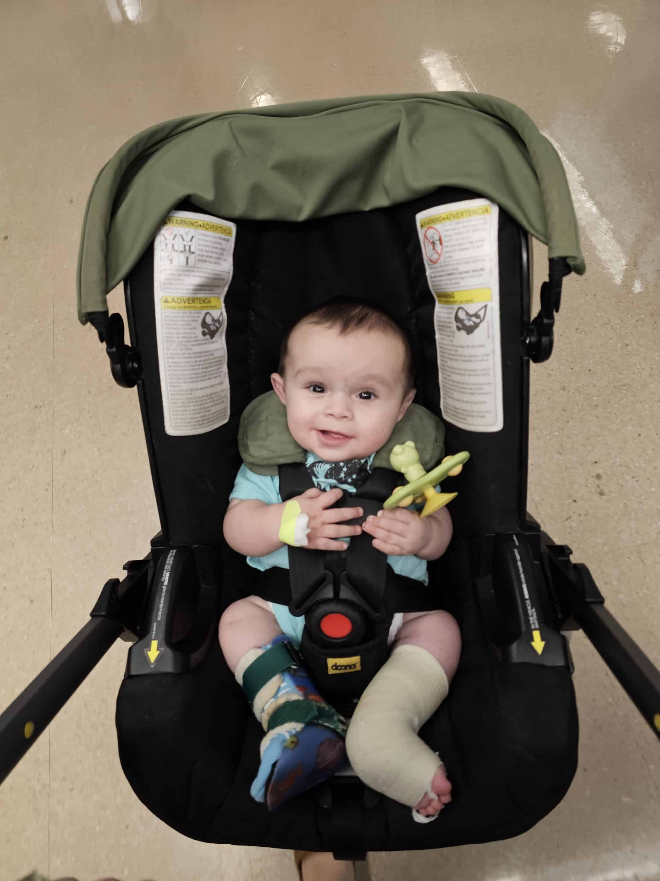
In serial casting, a cast is applied to the foot or feet once a week for three to five weeks. This brings the foot from upside down and turned inward position to a right side up and turned outward position. In some cases, a simple procedure under local anesthetic is performed to release tightness in the Achilles tendon and bring the foot into a normal position. After three to five weeks of progressive casting, a final cast is applied to hold the foot in the normal position for three weeks.

Infant Clubfoot Care: 2 months old to 7 to 12-months-old

After serial casting is completed, the next phase in clubfoot treatment is bracing to maintain the correction that was achieved through casting. Babies will wear a brace called a boot and bar brace consisting of soft silicone lined shoes with soft suede straps. These shoes are connected to each other by a bar to keep the feet turned outward.

Scottish Rite follows the protocol set by the International Clubfoot Congress of wearing the brace for 23 hours a day until the baby is beginning to pull up to stand, which usually happens between 7 to 12 months of age. Bracing does not interfere with the baby’s ability to reach developmental milestones, such as rolling over, sitting independently or crawling.

Toddler Clubfoot Care: 7 to 12 months old to 4 years old

After the baby begins pulling up to stand, wearing of the boot and bar brace is transitioned to nighttime only until the child reaches age 4. If bracing is not done in its entirety, the risk of clubfoot recurrence approaches 100%.

According to research at Scottish Rite, 70-80% of children will not require further treatment after the Ponseti method. Unfortunately, because clubfoot is a structural, congenital difference, approximately 20% of children will relapse and need further treatment, despite the medical providers and families doing everything correctly.

While clubfoot care may seem daunting, it is a safe and pain-free process that will give your baby the best outcome and prevent them from having difficulties later in life. Our team in the Center for Excellence in Foot stay with our patients and families every step of the way.

Learn more about clubfoot.

Infantile Clubfoot Deformity

Infantile Clubfoot Deformity

Clubfoot deformity occurs in one in 1,000 live births, making it the second most common congenital deformity of the foot (after postaxial polydactyly) and one of the most encountered infantile musculoskeletal congenital differences. In contrast to other common infantile foot deformities, such as metatarsus adductus and calcaneovalgus deformities, a clubfoot is not a normal foot that has been “packaged” into an abnormal position. Rather, a clubfoot is a true congenital deformity. There are structural differences inside the foot that create the outward deformity. The shape of the tarsal bones is not the same as that of a child with a normal foot. The elasticity of the soft tissues is much less than in a child with a normal foot, and arterial vessels are often absent or aberrant. Some of the extensor muscles in the leg have been shown to have an exponentially higher number of contractile elements (actin and myosin filaments) and are thus able to generate force at what is essentially a superhuman level.

Despite our understanding of the underlying anatomic and histologic differences in clubfeet, the exact etiology of the deformity remains unknown. Although genetic aberrancies have been identified that are associated with the clubfoot, these appear to be a result of somatic mutations, and thus, the deformity does not appear to run in families with any identifiable pattern of inheritance.

In approaching the treatment of a clubfoot, it is essential for families and providers to understand the presence of these congenital differences because our method of treating clubfoot cannot alter any of them. We can’t change the shape of the bones. We can’t improve the elasticity of the soft tissues. We can’t change the way the blood vessels move into the foot, and we can’t change the power with which certain muscles fire. Therefore, the goal of clubfoot treatment isn’t to create a normal foot. Rather, the goal of treatment is to take a structurally abnormal foot, which is also in an abnormal position, and through a series of casts applied weekly, slowly convert into an abnormal foot structurally that is ultimately in a near normal or completely normal position. Also, this method limits the need for surgery, which creates scarring and stiffness, thereby preserving as much motion of the foot as possible.

The Ponseti method of treatment is the gold standard of care worldwide for clubfoot deformity. This involves weekly serial casting of the involved foot or feet, with each cast applied in a manner to correct a different component of the clubfoot deformity. These casts are very effective at correcting every component of the clubfoot deformity with great success, save for the contracture of the Achilles tendon, which drives an oftentimes rigid equinus (plantarflexion) deformity of the ankle. Approximately 90% of clubfeet require a percutaneous transection of the Achilles tendon. This is performed in the clinic with topical anesthesia in children aged less than 3 months to overcome this component deformity. Following the heel cord tenotomy, the ankle can be acutely dorsiflexed to at least 10 to 15 degrees, and that position is maintained in a final cast that is kept in place for three weeks.

On average, it takes about four weeks of weekly casting to correct the component deformities of the cavus, hindfoot varus, and metatarsus adductus. That brings the foot from what is essentially an upside down and turned inward position to a right-side-up and turned-out position. At that point, if the ankle remains in equinus, this deformity is corrected with a heel cord tenotomy and a final three-week cast, resulting in a total time to achieve full correction of about seven weeks. As we ideally begin treatment within one or two weeks of life, most deformities are corrected by or before 2 months of age.

It cannot be overstated that deformity correction is just the first part of the treatment of a clubfoot deformity. Maintaining that correction is essential and, in many regards, more difficult. Seven weeks in a cast simply cannot overcome months of intrauterine gestation and the various congenital differences present within these feet. Without a means of maintaining correction, corrected clubfeet will universally and quickly revert to their initial position of deformity.
Maintenance of correction is achieved by keeping the ankles dorsiflexed and the feet externally rotated in what is typically called a “boot and bar” construct. While there are a variety of Ponseti-type braces available, most consist of soft silicone lined shoes with soft suede straps. Regardless of whether the deformity is unilateral or bilateral, these shoes are applied to both feet and connected to each other by a bar. The bar holds the shoes at 60 to 70 degrees of external rotation, and the shoe straps hold the ankles in neutral dorsiflexion.

Though bracing protocols vary to some degree, our protocol mirrors that agreed upon at the International Clubfoot Congress and consists of brace use for 23 hours a day until the child begins to pull to stand and 12 hours a night thereafter until age 4. The braces do not interfere with the achievement of developmental milestones, such as sitting up, rolling over, crawling or standing. The transition from full-time to part-time use is based entirely on the fact that once a child is able to pull to a standing position, they will soon spend more time on their feet. At that time, their own body weight will help to keep their feet flat on the ground. Until that time, babies spend most of their time lying down and sitting, during which the feet are held in a relaxed position. That can result in tightening of the heels cords and a recurrence of equinus.

Bracing is most difficult on parents for the first two weeks after the child comes out of their post-tenotomy cast. After seven weeks of casting, having anything other than cast padding surrounding their feet feels different and can cause fussiness. Parents are counseled extensively about this so they can anticipate and develop strategies to keep the braces in place despite frustration on the part of the child.

As mentioned, the use of the brace and the prescribed protocol is essential to long-term treatment success. If the braces aren’t used as recommended, the risk of recurrence of the deformity approaches 100%. These recurrences can be much more difficult to manage in older children than during infancy. It typically requires repeat casting and general anesthesia for either a repeat heel cord tenotomy or a more invasive surgical intervention.

Unfortunately, even when perfect correction is achieved with casting, and parents are compliant with brace use, a percentage of these deformities still recur and require additional treatment due to the structural congenital differences present. Research has demonstrated that with parental bracing compliance, about 80% of clubfoot patients never need any additional treatment. However, despite everyone’s best efforts, 20% of children still need some additional treatment due to recurrence. These recurrent deformities are most commonly the result of an imbalanced muscle pull across the foot due to a relatively overpowered tibialis anterior tendon. When relatively overpowered, this muscle, due to its insertion along the medial aspect of the foot, supinates the foot during dorsiflexion. That results in the touchdown of the lateral border of the foot during the initial stance phase of gait. This supination, in turn, slowly drives the foot inward and into further supination with weightbearing. Such recurrences are easily managed with a brief period of repeat casting to correct any relapsed deformities. That’s then followed by a transfer of the offending tendon to the dorsum of the foot to remove that supination moment during dorsiflexion.

With an experienced team of providers and parental dedication to brace use, these initially dramatic foot deformities can be corrected beautifully and without any long-term deficits or disabilities. However, it is important to educate parents early regarding certain differences in the foot or feet following treatment. A clubfoot is always smaller than a normal foot, sometimes half to a full shoe size smaller than an unaffected contralateral foot. In addition, the calf musculature of children with clubfoot tends to be smaller, though this size difference does not appear to affect strength. These differences are strictly cosmetic and do not alter function. The goal of clubfoot treatment is to allow a foot or feet to sit flat to the ground, have as much flexibility as possible, allow the child to wear any shoes they want, play any sports they want, have any job they want, and do whatever they want with the foot. In achieving this, their corrected clubfoot will function no differently than a normal foot and will never limit a child in any way in any activity they desire.

Download the PDF.

Dr. Anthony I. Riccio is a pediatric orthopedic surgeon and the Director of the Center for Excellence in Foot at Scottish Rite for Children.