When your young athlete has an injury or hurts, you want to know what happened and what comes next. Scottish Rite’s Sports Medicine team can help your injured athlete get back in the game safely.
Our Approach to Sports Medicine
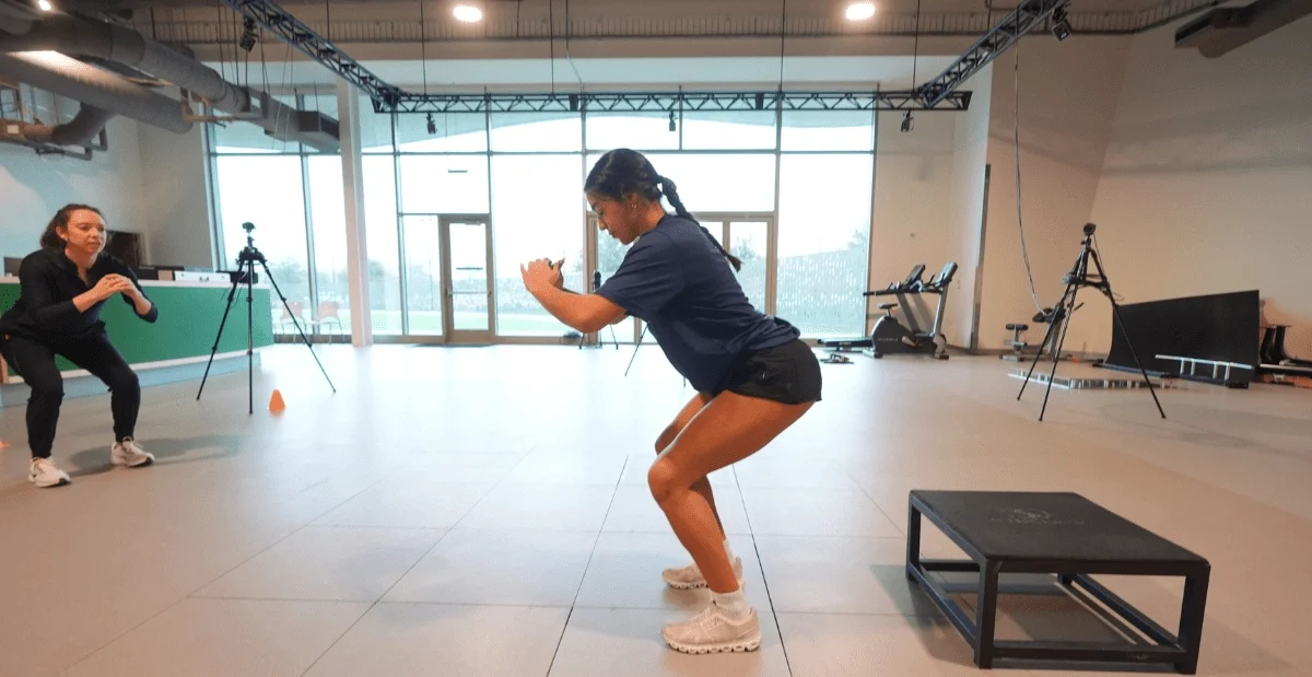
Whether your young athlete injured a knee at yesterday’s game or is experiencing joint pain, swelling or instability, our Sports Medicine team can help.
Early evaluation and treatment can often lead to faster recovery and may help avoid surgery.
At Scottish Rite, we understand the distinct needs of young and growing athletes. We treat more than the injury. We focus on the whole athlete, treating the injury while also supporting their goals to heal and safely return to play.
Our Sports Medicine team will focus on:
- Delivering the most advanced, research-based treatments
- Protecting their growth plates
- Supporting long-term development
- Preventing future injuries
- Supporting mindset and emotional recovery
- Considering factors that affect healing, including nutrition and sleep
Research that Improves Recovery
Our world-renowned Sports Medicine team has extensive experience in treating both common and complex sport-related injuries and conditions. They also lead and conduct nationally and internationally acclaimed research that has helped improve treatments for young athletes.
Your child’s treatment will be driven by research and results, all centered around their sport-specific needs and personal goals.
A Team in Your Corner
At Scottish Rite, your child will have access to a multidisciplinary team of sports medicine experts who will develop the best game plan for treatment, rehabilitation and a safe return to sport. Their care team may include:
- Sports medicine physicians who evaluate your child’s injury and manage conditions that do not require surgery.
- Pediatric orthopedic sport surgeons who treat complex joint and ligament injuries with advanced arthroscopic and surgical solutions.
- Athletic trainers who fit braces and assist in developing exercise programs unique to each condition
- Physical therapists who guide rehabilitation to help rebuild strength, range of motion and more after injury or before and after surgery
- Psychologists who offer support that addresses fear of re-injury, time away from sport and pain management techniques.
This team approach ensures that your child has coordinated, efficient care all in one location.
What to Expect with Sports Medicine
We understand the stress that sports injuries may present. We work hard to see athletes as quickly as possible, sometimes in the same day.
At their first appointment, we’ll take the time to understand your young athlete’s injury history and athletic goals. We’ll discuss your child’s:
- Symptoms and concerns
- Stage of growth and development
- Sport and their position
- Goals and competition level
We’ll also consider on how their injury is affecting their everyday life and function outside of their sport.
By the end of the appointment, we will help you and your child understand:
- What may be causing your child’s symptoms
- Next steps in their treatment plan
- What to expect for their recovery and return to play
In some cases, we may order additional imaging to better understand your child’s injury. This may include:
- EOS imaging, which is a low dose, standing X-ray that helps us to evaluate leg alignment
- Ultrasound to assess muscles, tendons and other soft tissues
- MRI for a more detailed view of ligaments, cartilage and other structures
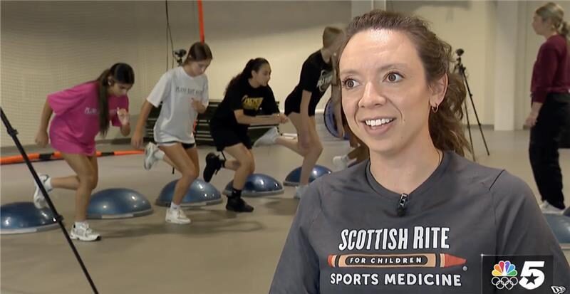
Support from Athletic Trainers
You’ll also meet with our athletic trainers, who are important members of your child’s care team.
Our athletic trainers are familiar with a wide range of sports, like baseball, football, gymnastics and dance. They bring years of sideline experience, understanding how injuries happen in real games and practices. Through our Sports Medicine clinic, they support young athletes with injuries.
Your child’s athletic trainer will assist with:
- Injury evaluations and progress
- Treatment education, including home exercise programs
- Brace or orthotic fitting
- Sport-specific return-to-play guidance
They are also available throughout your child’s treatment to answer questions and provide additional support.
Why Choose Us?
Timely, thoughtful care: We know sports injuries need attention. Our Sports Medicine team provides timely treatment so your young athlete can start recovering as quickly as possible.
Innovation through research: Our Sports Medicine team leads and publishes groundbreaking research that improves patient outcomes, reduces complications during surgery and advances the practice of pediatric sports medicine worldwide.
Built for growing athletes: Children are not small adults. Whether your child has a soccer injury, concussion or a rare cartilage disorder, we’ll develop a customized treatment plan. This ensures we protect their long-term growth, development and performance.
-
We aim to see patients as soon as possible. In some cases, we can schedule an appointment within one business day.
-
You can schedule an appointment by calling us directly at 469-515-7100 or requesting online. Our Sports Medicine team can guide you through scheduling.
-
No, your child does not need a referral to make an appointment with our sports medicine experts.
-
We have extensive expertise in treating children and teens who participate in a wide range of sports, including:
- Baseball and T-ball
- Basketball
- Cheer and tumbling
- Climbing
- Cross country and running
- Dance, drill team and ballet
- Diving
- Equestrian activities
- Figure skating
- Football
- Golf
- Gymnastics
- Ice hockey
- Lacrosse
- Martial arts
- Motorcross
- Pickleball
- Rodeo
- Rugby
- Soccer
- Softball
- Swimming
- Tennis
- Track and field
- Volleyball
- Wrestling